Corneal endothelial cell therapy replaces the failing endothelium with cultured cells, instead of transplanting a full donor graft.
Cells from one donor cornea can in theory treat many patients, which would ease the global shortage of tissue.
The first clinical results, from Japan, have been encouraging (see eye banking and the donor cornea).
The Problem Corneal Endothelial Cell Therapy Addresses
The endothelium pumps fluid out of the cornea, and its cells do not divide in adults.
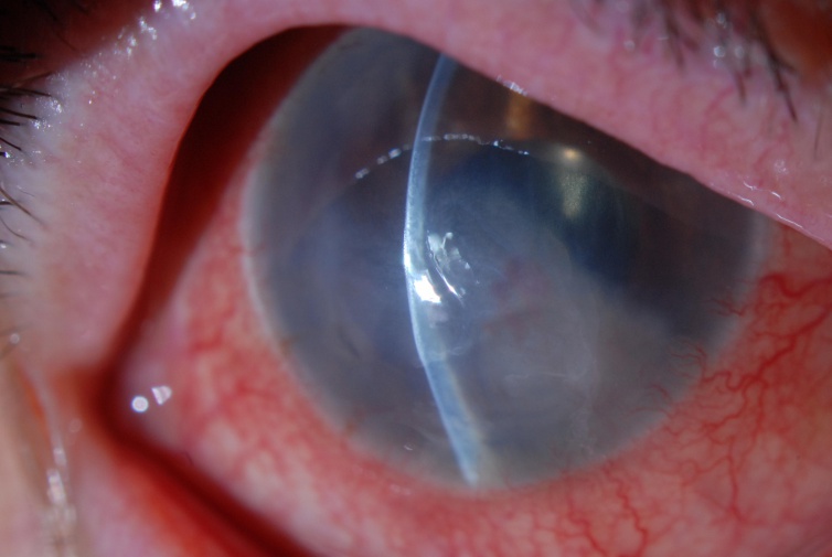
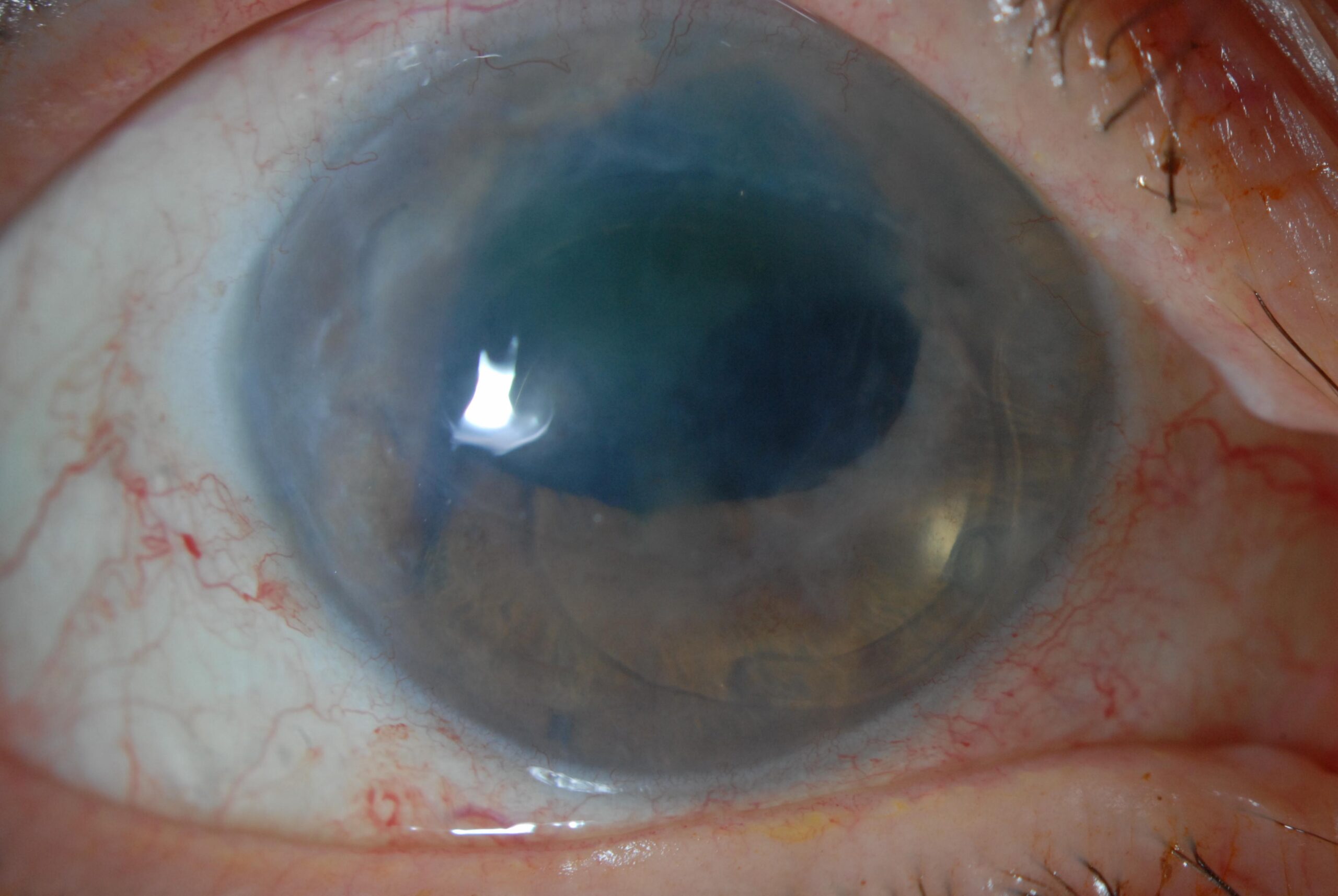
When Fuchs dystrophy, surgery, or injury destroys too many, the cornea swells and vision falls (see corneal endothelial decompensation).
Endothelial keratoplasty restores clarity, but needs donor tissue, and one cornea serves one patient (see DSEK and DMEK).
Corneal Endothelial Cell Therapy: The Idea
Laboratories can grow endothelial cells from a donor cornea, expand them, and inject them into the anterior chamber of a patient.
The cells attach to the back of the cornea and form a new monolayer, if the technique helps them stick.
All-fit Slit-Lamp Adapter
Record and share exactly what you see at the slit lamp. One adapter fits any slit lamp or surgical microscope — and any smartphone.
From Choroida — the team behind this siteThe Role of ROCK Inhibitors
Rho kinase inhibitors improve the adhesion and survival of endothelial cells and promote their migration.
In the injection technique, the surgeon gives cultured cells together with a ROCK inhibitor, and the patient lies face-down for a few hours so that cells settle on the cornea (see the references).
Corneal Endothelial Cell Therapy: The Japanese Trials
A clinical study of eleven patients with bullous keratopathy injected cultured human corneal endothelial cells with a ROCK inhibitor.
- The corneas became clear in nearly all patients, with a return of normal thickness within weeks (see the references)
- Endothelial cell density rose to levels that supported clarity
- Investigators reported no serious adverse effects, and follow-up of five years showed durable benefit in most (see the references)
Japan approved a product based on this approach in 2023.
Other Strategies
- Injection of ROCK inhibitor alone after removal of diseased endothelium, which may let healthy peripheral cells migrate and cover the center (see Descemet stripping only)
- Cell sheets or carriers that deliver cells to the back of the cornea
- Cells derived from stem cells, to avoid the need for donor corneas
- Gene and drug therapy that protects endothelial cells in Fuchs dystrophy (see Fuchs endothelial dystrophy)
Corneal Endothelial Cell Therapy: Advantages
- One donor cornea can supply many doses
- Less surgery, with an injection and positioning
- Potentially lower rejection risk, though immune reactions are possible
- A way to reach patients in areas with a shortage of tissue
Limits and Questions
- Investigators still follow the long-term survival of the injected cells
- Manufacturing needs strict standards and cost control
- Patients with severe damage or scarring may need a graft
- Evidence outside Japan is still limited, and trials are under way in other regions
Corneal Endothelial Cell Therapy: Counselling
Patients should know that the treatment is new, that it is available in a few centers or in trials, and that standard endothelial keratoplasty remains an effective option.
Example Patient
A 67-year-old man with bullous keratopathy after cataract surgery has a swollen cornea and hand-motion vision.
In a trial, he receives an injection of cultured endothelial cells with a ROCK inhibitor, and lies face-down for three hours.
Over the next weeks the cornea clears, and the pachymetry falls, which is the pattern described in the first studies.
Questions About the Source of Cells
Cells come from screened donor corneas, expanded under strict manufacturing conditions.
Because they are not matched to the patient, rejection is possible, so topical steroids and follow-up are needed.
Outlook
If the results of early studies are repeated elsewhere, a single donor cornea might treat many patients, which would help regions with long waiting lists.
Clinicians should watch for trials and approvals in their country.
Practical Messages for Clinics
Document endothelial cell counts and corneal thickness in patients with early failure, since they may become candidates for cell therapy.
Keep records that help patients to qualify for trials, and refer to centers that offer these studies.
Safety and Regulation
Cell products must meet strict standards for sterility, identity, and potency, and laboratories test each batch.
Regulators ask for long-term follow-up, because cells could in theory change over time.
So far, trials report a good safety record, with rare cases of raised pressure or inflammation that responded to treatment.
Questions Patients Ask
Patients ask whether the injection means they will never need a transplant, and the honest answer is that the long-term data are still being collected.
They also ask how long the cornea takes to clear, which in the first studies was a matter of weeks, with a period of positioning and drops.
For the latest studies, see the PubMed results for corneal endothelial cell therapy.


Document what you see
Two smartphone imaging tools built for everyday clinic use — one for the slit lamp, one for the fundus.
From Choroida — the team behind this siteReferences
- Kinoshita S, Koizumi N, Ueno M, et al. Injection of cultured cells with a ROCK inhibitor for bullous keratopathy. N Engl J Med. 2018;378:995-1003.
- Numa K, Imai K, Ueno M, et al. Five-year follow-up of first 11 patients undergoing injection of cultured corneal endothelial cells for corneal endothelial failure. Ophthalmology. 2021;128:504-514.
- Okumura N, Koizumi N, Ueno M, et al. Rho kinase inhibitor enables cell-based therapy for corneal endothelial dysfunction. Sci Rep. 2016;6:26113.
- American Academy of Ophthalmology. Basic and Clinical Science Course, Section 8: External Disease and Cornea. AAO; current edition.

Writes and reviews the clinical articles on Educate. Every post is checked against peer-reviewed sources before it goes live.