An implantable collamer lens is a thin, soft lens placed inside the eye, in front of the natural lens, to correct refractive error.
It is used mainly for moderate and high myopia, in patients whose corneas are too thin or too irregular for laser treatment.
It preserves the cornea, and it is reversible, but it is an intraocular operation with its own risks.
What Is an Implantable Collamer Lens?
The lens is made of collamer, a flexible copolymer of collagen and a hydrophilic polymer that the eye tolerates well.
The surgeon also folds it and injects it through a small corneal incision into the posterior chamber, where the haptics rest in the ciliary sulcus.
Newer designs have a small central port, which allows aqueous to flow through the lens, so that a preoperative iridotomy is not needed (see the references).
In addition, toric versions correct astigmatism, and the lens is available in a range of powers for myopia, and in some regions for hyperopia.
Who Is a Good Candidate?
- Age usually between 21 and 45, with a stable refraction
- Moderate to high myopia, commonly from about minus 3 to minus 18 diopters
- A thin or irregular cornea, high myopia beyond the range of laser, or dry eye that makes laser a poor choice
- Adequate anterior chamber depth, usually 3.0 millimeters or more, and a healthy endothelium with a cell density above about 2,300 to 2,500
- No history of glaucoma, uveitis, or cataract
Those who do not qualify include patients with a shallow chamber, an unhealthy endothelium, or zonular problems.
All-fit Slit-Lamp Adapter
Record and share exactly what you see at the slit lamp. One adapter fits any slit lamp or surgical microscope — and any smartphone.
From Choroida — the team behind this siteThe Operation
- Preoperative measurements include the white-to-white distance, the sulcus size, and the chamber depth, to select the length of the lens
- The procedure takes about 15 minutes, with topical or local anesthesia and a small incision
- The surgeon places the lens behind the iris, with the pupil dilated, and removes the viscoelastic
- Patients usually return to normal activities in a few days
Clinicians may do both eyes on the same day or a few days apart.
Results of Implantable Collamer Lens
Studies show high rates of good uncorrected vision and high satisfaction.
A quality of vision that is often better than after laser for high myopia (see the references).
The loss of best-corrected vision is rare, and many patients gain lines, since the lens produces less minification than glasses.
Results are stable in most patients, and surgeons can remove the lens if needed.
The Importance of the Vault
The vault is the distance between the back of the lens and the front of the natural lens.
- Ideal values are about 250 to 750 micrometers, or about half to one and a half times the corneal thickness
- A low vault increases the risk of contact with the natural lens, and so of cataract
- A high vault can also narrow the angle and raise the pressure
Also, measurement of the vault by anterior segment OCT or ultrasound biomicroscopy at follow-up helps detect problems (see ultrasound biomicroscopy).
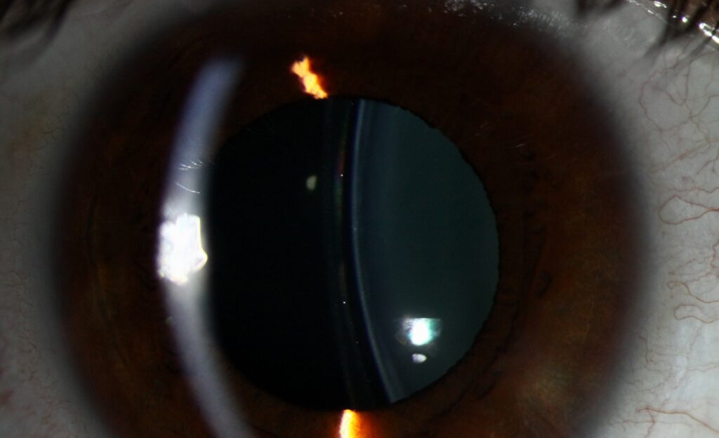
Complications of Implantable Collamer Lens
- Anterior subcapsular cataract: a known risk, usually due to contact with the lens or surgical trauma, with an incidence of a few percent over years
- Raised intraocular pressure: from retained viscoelastic, steroid response, pupillary block in lenses without a port, or a high vault
- Endothelial cell loss, which should also be monitored with specular microscopy (see specular microscopy)
- Pigment dispersion and glare
- Rotation of toric lenses, and an incorrect lens size
- Rare infection and retinal detachment, the latter related to myopia
Regular follow-up is needed for life.
Compared With Laser Vision Correction
Laser removes corneal tissue and may cause dry eye and ectasia in thin corneas, while the lens adds a device inside the eye but leaves the cornea intact (see LASIK).
In high myopia, the lens usually gives better vision quality, while laser is simpler and less costly for low to moderate errors.
Counselling
Explain the benefits and the risks, such as cataract in later life, and the need for regular checks.
Also, discuss expectations about night vision, and the cost.
Implantable Collamer Lens: Follow-Up Schedule
Review the patient on the first day, at one week, at one month, and at three months, with pressure, vault, and lens position checked, and then yearly with endothelial counts and lens clarity.
Patients should avoid eye rubbing, report sudden pain or vision loss at once, and keep yearly visits for life, since a late cataract or pressure change can be treated well when found early.


Document what you see
Two smartphone imaging tools built for everyday clinic use — one for the slit lamp, one for the fundus.
From Choroida — the team behind this siteReferences
- Sanders DR, Doney K, Poco M; ICL in Treatment of Myopia Study Group. United States Food and Drug Administration clinical trial of the Implantable Collamer Lens (ICL) for moderate to high myopia: three-year follow-up. Ophthalmology. 2004;111:1683-1692.
- Packer M. The Implantable Collamer Lens with a central port: review of the literature. Clin Ophthalmol. 2018;12:2427-2438.
- Kamiya K, Shimizu K, Igarashi A, et al. Posterior chamber phakic intraocular lens implantation: comparative, multicentre study in 351 eyes with low-to-moderate or high myopia. Br J Ophthalmol. 2018;102:177-181.
- American Academy of Ophthalmology. Basic and Clinical Science Course, Section 13: Refractive Surgery. AAO; current edition.

Writes and reviews the clinical articles on Educate. Every post is checked against peer-reviewed sources before it goes live.