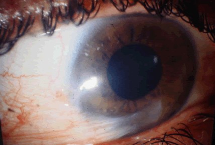
Pterygium recurrence is the regrowth of fibrovascular tissue onto the cornea after surgical removal.
It was the great problem of pterygium surgery for decades, and it still occurs, although modern techniques have brought the rate down greatly.
The recurrent lesion is often more aggressive than the original, so prevention is important.
What Is Pterygium Recurrence?
A pterygium is a fibrovascular growth of the conjunctiva that extends onto the cornea, driven by ultraviolet exposure and chronic irritation (see pterygium).
After excision, the exposed sclera and the limbus are at risk of fibroblast proliferation and neovascularization, and the lesion can grow back from the limbus.
Recurrence is defined as fibrovascular tissue that crosses the limbus onto the cornea after surgery, usually within the first six months, and most within a year.
Recurrences that appear later often reflect continuing exposure or other disease of the ocular surface.
How Common Is It?
The rate depends on the surgical technique.
- Bare sclera excision: recurrence rates are high, reported at roughly 30 to 80 percent in various series
- Conjunctival autograft: the rate falls to about 5 to 10 percent or lower in many series, especially with good technique
- Amniotic membrane graft: intermediate, with rates that vary between studies
- Adjunctive mitomycin C: lowers recurrence in bare sclera and in graft procedures, but with a risk of scleral melting
A review of pterygium treatment by Hirst concluded that conjunctival autograft, with or without fibrin glue, offered the best balance of recurrence rate and safety.
All-fit Slit-Lamp Adapter
Record and share exactly what you see at the slit lamp. One adapter fits any slit lamp or surgical microscope — and any smartphone.
From Choroida — the team behind this siteRisk Factors of Pterygium Recurrence
- Young age, in particular under 40
- Fleshy, thick, and highly vascular lesions
- Large lesions and those with a high degree of inflammation
- Continued exposure to sunlight, dust, wind, and dry conditions
- A history of recurrence
- Poor technique, such as leaving Tenon tissue, a small graft, or exposed sclera
- Ocular surface disease such as dry eye
Prevention at the Time of Surgery
- Excise the pterygium head and body, and remove the abnormal subconjunctival Tenon tissue
- Cover the bare sclera with a conjunctival autograft, taken from the superior conjunctiva, with the correct orientation (see conjunctival autograft pterygium surgery)
- Use fibrin glue or sutures to fix the graft in place, with minimal inflammation
- Consider intraoperative mitomycin C at 0.02 percent for a short time in high-risk eyes, with caution regarding the sclera
- Avoid cautery to the sclera that could damage the tissue
- Control inflammation after surgery with topical steroids, with a slow taper
Postoperative sunglasses and lubricants support healing and reduce the stimulus for recurrence.
Treatment of a Recurrent Pterygium
- Observation: a small, quiet recurrence that does not threaten the visual axis can be observed
- Repeat excision with autograft: the standard approach, with removal of scar tissue and a larger graft
- Amniotic membrane or limbal-conjunctival graft if conjunctiva is limited (see amniotic membrane transplantation)
- Adjuvants: mitomycin C or anti-VEGF drops or injections are used by some surgeons, although the evidence is limited
- Beta radiation: used in the past, but associated with severe late complications, so it is avoided
Treat dry eye and inflammation, and reassess the technique used in the first operation.
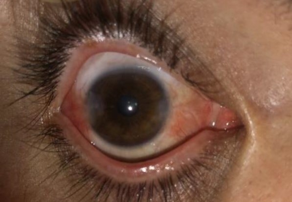
Pterygium Recurrence: Complications of Treatment
- Scleral melting, necrosis, and perforation, associated with mitomycin C and radiation
- Graft retraction, necrosis, and granuloma
- Corneal scarring and astigmatism
- Dellen and infection
Patients should be told about these risks and instructed to return if there is pain, redness, or a change in vision.
Counselling
Explain that the lesion can recur but that careful surgery lowers the chance, and that protecting the eye from sunlight reduces the stimulus.
Ask the patient to wear sunglasses and a hat outdoors and to use lubricants for dryness.
Practical Surgical Pearls
- Use a graft that is slightly larger than the defect, since it tends to shrink
- Avoid placing the graft with the epithelial side down
- Remove all Tenon tissue from the graft and the bed, since it is a source of fibrovascular proliferation
- Keep the limbus clear, with the graft edge in contact with the limbus
- Avoid excessive tension and avoid leaving a gap between the graft and the surrounding conjunctiva
Fibrin glue shortens operating time and reduces postoperative discomfort, and it lowers the risk of suture-related inflammation.
Observation After Surgery
Review the patient at one day, one week, one month, and three months, and look for early signs of recurrence such as new injection and vessel growth toward the cornea.
Early recurrence may respond to topical steroids or other anti-inflammatory therapy before it crosses the limbus.
Advise the patient to report redness, a rising lesion, or a change in vision.
Record the grade of the lesion and the technique used, so that the next surgeon can learn from earlier treatment.
For the latest studies, see the PubMed results for pterygium recurrence.


Document what you see
Two smartphone imaging tools built for everyday clinic use — one for the slit lamp, one for the fundus.
From Choroida — the team behind this siteReferences
- Hirst LW. The treatment of pterygium. Surv Ophthalmol. 2003;48:145-180.
- Kenyon KR, Wagoner MD, Hettinger ME. Conjunctival autograft transplantation for advanced and recurrent pterygium. Ophthalmology. 1985;92:1461-1470.
- Mannis MJ, Holland EJ, eds. Cornea. 4th ed. Elsevier; 2017.
- American Academy of Ophthalmology. Basic and Clinical Science Course, Section 8: External Disease and Cornea. AAO; current edition.

Writes and reviews the clinical articles on Educate. Every post is checked against peer-reviewed sources before it goes live.