The clear corneal incision is the standard entry for cataract surgery, and its construction determines whether the wound seals without sutures and stays sterile.
A well-made incision is also self-sealing, resists intraocular pressure, and causes little astigmatism.
A poor one allows leakage, hypotony, and a path for infection.
Why Clear Corneal Incision Construction Matters
The wound must stay closed under the effect of the intraocular pressure, eye rubbing, and blinking.
In addition, the inner lip is pushed against the outer lip by pressure, which acts as a valve in a well-constructed tunnel.
A short, steep, or irregular incision does not seal well and leaks.
Studies showed that wounds with a short tunnel or a wound that gapes when the pressure fluctuates are more likely to allow fluid and microorganisms in.
Clear Corneal Incision: Architecture of the Wound
- Tunnel length: typically about 1.5 to 2 millimeters, with the length at least equal to the width of the wound
- Width: matched to the instrument and lens, usually 2.2 to 3.0 millimeters
- Location: temporal or on the steep axis, depending on the astigmatism
- Plane: a single plane or a two-plane design, with a stromal tunnel beneath the surface
- Internal opening: with a smooth edge in Descemet membrane
A square incision, or one that is slightly longer than it is wide, is more stable than a wide, short one.
The knife should be sharp, and the entry perpendicular at first and then flattened to create the tunnel.
All-fit Slit-Lamp Adapter
Record and share exactly what you see at the slit lamp. One adapter fits any slit lamp or surgical microscope — and any smartphone.
From Choroida — the team behind this siteSealing and Stromal Hydration
At the end of surgery, the surgeon hydrates the walls of the incision by injecting balanced salt solution into the stroma on both sides, which swells the tissue and closes the wound.
Also, the stromal hydration is reversible, and it does not replace a well-constructed wound.
The eye is then pressurized and the wound is checked for leakage.
Clear Corneal Incision: Testing for Leaks
- Dry the wound surface and apply gentle pressure with a sponge
- Observe whether fluid leaks out of the wound
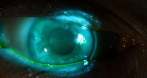
- Perform a Seidel test with fluorescein dye, which shows the stream of aqueous
- Check the pressure and the anterior chamber depth
A leaking wound should be sutured or repaired, as the risk of infection is higher.
Also, many surgeons use a single suture in any wound that does not hold.
Clear Corneal Incision: Wound Leaks After Surgery
A leak can occur in the first days if the wound is not sealed.
The patient may report a wet feeling, pain, or a shallow chamber.
Treat a small leak with a bandage lens and aqueous suppressants, and repair larger leaks with sutures (see corneal wound dehiscence).
In addition, hypotony may cause choroidal detachment, corneal folds, and endophthalmitis.
Infection Risk
A systematic review found a higher risk of endophthalmitis after clear corneal incisions than after scleral tunnel incisions, although recent practice improvements have narrowed the difference.
Other risk factors include wound leaks, posterior capsule rupture, and intraoperative events (see acute postoperative endophthalmitis).
Also, measures to lower the risk include meticulous wound construction, povidone-iodine antisepsis, and intracameral antibiotics (see intracameral antibiotics).
Astigmatism and Placement
The incision flattens the meridian where it is placed, and a larger or more central incision causes more astigmatism.
Place the incision on the steep meridian for a mild effect, and consider arcuate incisions for larger astigmatism.
A temporal approach reduces the risk of against-the-rule shift in many patients.
Practical Pearls
Make sure the speculum does not distort the cornea during wound construction.
Avoid thermal injury to the incision by keeping the irrigation flowing and the phaco tip clean.
Check the wound at the end, and do not hesitate to place a suture.
Instruments and Technique
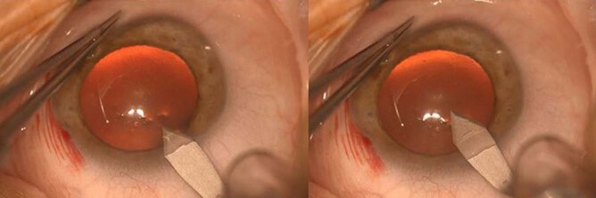
Use a sharp, single-use keratome of the right width, and check the blade before use.
Enter the cornea in a controlled manner, with the blade flat, advance it in the stroma to form the tunnel, and then angle it into the chamber.
A premarked or guarded blade helps to control the length of the tunnel, and trainees benefit from practicing on a model.
Avoid excessive manipulation of the wound with instruments, since stress can distort the architecture and thermal injury can shrink the tissue.
Wound Healing and Late Problems
After surgery, the wound gradually heals, and the stromal edema subsides within hours to days.
A small gaping on the inside may occur in the first days, and it usually resolves.
Late problems include epithelial ingrowth along the wound, scarring, and dellen near the incision, and these are uncommon.
Patient Instructions
Tell patients not to rub the eye, to avoid swimming and pressure on the eye, and to use the shield at night for the advised time.
They should report pain, redness, a wet feeling, or a sudden fall in vision, because these can be signs of a leak or an infection.
Teach them how to instill the drops without touching the eye with the bottle tip.


Document what you see
Two smartphone imaging tools built for everyday clinic use — one for the slit lamp, one for the fundus.
From Choroida — the team behind this siteReferences
- Masket S, Belani S. Proper wound construction to prevent short-term ocular hypotony after clear corneal incision cataract surgery. J Cataract Refract Surg. 2007;33:383-386.
- Taban M, Behrens A, Newcomb RL, et al. Acute endophthalmitis following cataract surgery: a systematic review of the literature. Arch Ophthalmol. 2005;123:613-620.
- Salmon JF. Kanski’s Clinical Ophthalmology: A Systematic Approach. 9th ed. Elsevier; 2020.
- American Academy of Ophthalmology. Basic and Clinical Science Course, Section 11: Lens and Cataract. AAO; current edition.

Writes and reviews the clinical articles on Educate. Every post is checked against peer-reviewed sources before it goes live.