
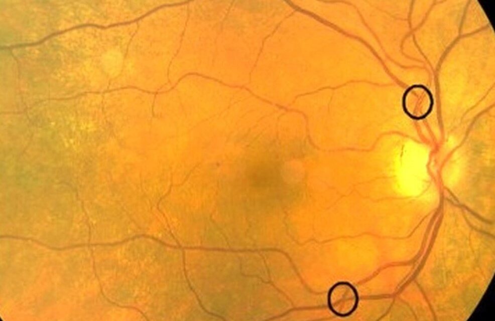
At an arteriovenous crossing, the artery and vein share a common adventitial sheath.
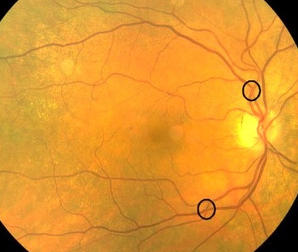
When the artery is hardened by long-standing hypertension, it presses on the vein beneath or above it, and the vein changes shape in a predictable way.
Three eponymous signs describe those changes, and they are often quoted together without being clearly separated.
The three signs
Gunn sign
The vein appears to taper on both sides of the crossing, as though it were being pinched.
This is the classic AV nicking.
The column of blood seems to disappear beneath the artery, a finding which gets more obvious as the artery wall thickens.
Salus sign
The vein is deflected at the crossing.
Instead of running straight, it bends, forming an S or a shallow curve.
The deflection usually becomes visible when the vein crosses beneath an arteriole that has lost its elasticity.
Bonnet sign
The vein is dilated on the distal side of the crossing, a banking, as if the flow were being held back.
It points to venous outflow obstruction at the crossing, and it is the sign most closely linked with a branch vein occlusion at that site.
Why they matter
They mark chronic arteriolosclerosis, which implies sustained hypertension over years, not a recent blood pressure spike.
That is useful because the same eye may show only a few findings while the patient has had hypertension for a long time.
They also explain why branch retinal vein occlusion occurs at crossing points.
Compression increases turbulence and endothelial damage, and a thrombus forms (see branch retinal vein occlusion).
How they fit into grading
The older Keith-Wagener-Barker and Scheie classifications used these signs to separate mild from moderate disease.
Current practice puts less weight on grading and more on the question of whether there is target-organ damage, but AV crossing changes still help with risk stratification (see hypertensive retinopathy).
Examination tips
- Look at the superotemporal and inferotemporal arcades first, where crossings are numerous
- Use a green filter to improve contrast of the vessel walls
- Document with photographs, since observer agreement on mild nicking is poor
- Do not call an isolated nick significant; look for the whole picture, including arteriolar narrowing and wire changes
Differentiating them from physiological crossings
Normal crossings are common and most do not show any change.
A vein passing over an artery should keep its caliber and direction.
Concealment of the vein under the artery, if the vein is seen to vanish and reappear, is another way of describing Gunn’s tapering.
In children and young adults a mild tortuosity at crossings is physiological, so the signs carry meaning only in the setting of arteriolar narrowing, a broadened light reflex, or a risk profile that fits.
Photographic studies, including the Atherosclerosis Risk in Communities study, found that AV nicking is associated with long-term blood pressure and with stroke risk, even after adjusting for current blood pressure.
What to do with the finding
- Record the grade of hypertensive change and the number of crossings affected in each eye
- Check for associated signs: arteriolar narrowing, hemorrhages, cotton wool spots, disc swelling
- Send a letter to the primary physician when the signs are new, so that blood pressure and cardiovascular risk get a formal review
- Ask patients with a branch vein occlusion about hypertension, since the occlusion may be the first sign of undiagnosed disease
Management
There is no ocular treatment.
The task is blood pressure control, cardiovascular risk assessment, and referral when there are other signs of end-organ damage.
Why the vein is the one that changes
At a crossing, the artery and the vein share a common adventitial sheath, so the thickened arteriolar wall presses directly on a thin-walled vein. The vein cannot widen sideways, so the column narrows on both sides of the crossing, deviates, or dilates beyond it. When the artery crosses over the vein, as it does in most crossings, the effect is more obvious than when the vein lies on top.
Practical points at the ophthalmoscope
Examine the crossings close to the disc first, where the vessels are larger and the signs are easier to see. Mild nicking can be normal in older patients, so look for several crossings that show the same change, and for other signs of arteriolar disease. In a patient with a branch vein occlusion, check the fellow eye for nicking, since a tendency to occlusion at crossings is often bilateral.
Record the findings with the patient's blood pressure and the date, so that later examiners can judge whether the changes are stable.


Document what you see
Two smartphone imaging tools built for everyday clinic use — one for the slit lamp, one for the fundus.
From Choroida — the team behind this siteReferences
- Wong TY, Mitchell P. Hypertensive retinopathy. N Engl J Med. 2004;351:2310-2317.
- Scheie HG. Evaluation of ophthalmoscopic changes of hypertension and arteriolar sclerosis. Arch Ophthalmol. 1953;49:117-138.
- Klein R, Klein BE, Moss SE, Wang Q. Hypertension and retinopathy, arteriolar narrowing, and arteriovenous nicking in a population. Arch Ophthalmol. 1994;112:92-98.
- Hubbard LD, Brothers RJ, King WN, et al. Methods for evaluation of retinal microvascular abnormalities associated with hypertension/sclerosis in the Atherosclerosis Risk in Communities Study. Ophthalmology. 1999;106:2269-2280.
Test yourself
A few questions straight from this article.
-
Which retinal arteriovenous crossing change defines the Gunn sign?
In the Gunn sign the vein appears to taper on both sides of the crossing as though pinched; this is the classic arteriovenous nicking. -
Which retinal arteriovenous crossing change defines the Salus sign?
In the Salus sign the vein is deflected at the crossing, bending into an S or shallow curve instead of running straight. -
Which retinal arteriovenous crossing change defines the Bonnet sign?
The Bonnet sign is dilatation, or banking, of the vein on the distal side of the crossing, pointing to venous outflow obstruction at that site. -
How does compression at an arteriovenous crossing lead to branch retinal vein occlusion?
Compression at the crossing increases turbulence and endothelial damage, and a thrombus then forms, which is why branch vein occlusions occur at crossing points. -
Why is it the vein, rather than the artery, that changes shape at a crossing?
Artery and vein share a common adventitial sheath, so a thickened arteriolar wall presses directly on the thin-walled vein, which cannot widen sideways. -
What do arteriovenous crossing signs tell you about a patient's hypertension?
These signs mark chronic arteriolosclerosis, which implies sustained hypertension over years rather than a recent blood pressure spike. -
Why should arteriovenous crossing changes be documented photographically?
Documentation with photographs is advised because observer agreement on mild nicking is poor, making a recorded image more reliable than description alone. -
In children and young adults, what is the significance of mild tortuosity at arteriovenous crossings?
Mild tortuosity at crossings is physiological in children and young adults, so the signs carry meaning only alongside arteriolar narrowing, a broadened light reflex or a fitting risk profile. -
What is the management of arteriovenous crossing signs found on fundus examination?
There is no ocular treatment; the task is blood pressure control, cardiovascular risk assessment, and referral when there are other signs of end-organ damage. -
Photographic studies have linked arteriovenous nicking to which outcome, independent of current blood pressure?
Photographic studies, including the Atherosclerosis Risk in Communities study, linked arteriovenous nicking to long-term blood pressure and to stroke risk even after adjusting for current blood pressure.
