Vitrectomy complications range from common and benign changes, such as cataract after the first year, to rare and sight-threatening events such as endophthalmitis.
Modern small-gauge systems have reduced operating time and inflammation, but they have not removed risks.
A clear picture of what to expect at each stage of the postoperative course helps the clinician to react early.
Vitrectomy Complications by Timing
It is useful to divide complications into those that arise during the operation, in the first days, in the first weeks, and later.
Intraoperative events include iatrogenic breaks, lens touch, suprachoroidal hemorrhage, and vitreous incarceration at sclerotomies.
Early postoperative problems include raised pressure, hypotony, fibrin reaction, and endophthalmitis.
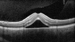
Later problems include cataract, retinal detachment, epiretinal membrane, and macular edema (see pars plana vitrectomy).
Each stage has typical presentations, and the symptoms often point directly to the cause.
Common Complications
- Cataract: nuclear sclerosis progresses in most phakic eyes within one to two years, particularly in patients older than 50 and after gas tamponade
- Raised intraocular pressure: from gas expansion, silicone oil, inflammation, or steroid response
- Iatrogenic retinal breaks: formed at sclerotomies or during peeling of membranes, sometimes found only after surgery
- Postoperative retinal detachment: from missed or new breaks, or from proliferative vitreoretinopathy (see proliferative vitreoretinopathy)
- Vitreous hemorrhage: particularly in diabetic eyes, from fibrovascular tissue or sclerotomy sites
- Hypotony: from wound leak or ciliary body shutdown, with choroidal detachment in severe cases
Fundus Explorer Pro
Photograph the retinal findings described here with the phone already in your pocket — 22 D optics and built-in illumination in one handheld unit.
From Choroida — the team behind this siteLess Common but Serious Events
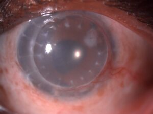
Endophthalmitis occurs in a small proportion of cases, and it presents with pain, loss of vision, and a hypopyon in the first days (see acute postoperative endophthalmitis).
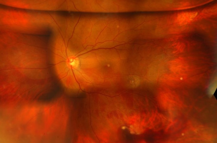
Suprachoroidal hemorrhage is rare and causes sudden pain and a dark, elevated choroidal mass, and it requires urgent attention.
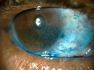
Corneal complications include epithelial defect, edema, and band keratopathy, especially with silicone oil in contact with the endothelium.
Macular phototoxicity from the endoilluminator can occur with prolonged high-intensity light.
Gas-related events include a rise in pressure during air travel or at altitude, and nitrous oxide anesthesia can cause expansion in an eye with intraocular gas.
Evaluation of a Problem After Surgery
Measure acuity, pressure, and the state of the wounds, and examine the anterior chamber, the lens, and the posterior segment with a dilated pupil.
Look for fibrin, hypopyon, corneal edema, and gas or oil position.
Use OCT of the macula when the acuity is lower than expected, and use B-scan ultrasonography if the view is poor (see B-scan ultrasonography).
A patient with pain and falling vision in the first week needs urgent assessment for endophthalmitis.
Management
Pressure rises are usually managed with topical agents, and gas can be partly released in severe cases.
Endophthalmitis requires vitreous tap and intravitreal antibiotics, with repeat vitrectomy if the eye is not responding.
A retinal redetachment needs prompt reoperation, and the choice of tamponade depends on the cause.
Cataract surgery is straightforward in many eyes after vitrectomy, but it can be technically harder because of the lack of vitreous support, and the zonules may be weak.
Patients should be warned about the lens changes, the need for positioning, and the restrictions on flying, high altitude, and nitrous oxide.
Careful written instructions and an easy way to reach the clinic help to catch problems early.
Reducing the Risk
Many complications can be prevented or softened by planning.
Careful preoperative examination identifies zonular weakness and corneal problems, and a discussion with the patient about the lens sets expectations.
During the procedure, meticulous shaving of the vitreous base, indentation of the periphery, and inspection for breaks at the end of the case reduce the rate of iatrogenic tears.
Controlling infusion pressure and avoiding prolonged high-intensity illumination protect the retina and the macula.
After surgery, early review at day one and again within a week detects pressure spikes, wound leaks, and early infection.
Follow-Up Schedule
A typical pattern is review on the first day, at one week, and at one month, with later visits adjusted to the disease treated and the tamponade used.
Patients with gas should avoid altitude and nitrous oxide until the bubble has gone, and should carry a wristband or card that says so.
Document the lens status, the pressure, and the tamponade at each visit, because they guide the next decision.
Keep a record of the gauge used, the tamponade, the retinopexy, and any intraoperative event in the operative note, because this information changes how a postoperative problem is interpreted.
When something unexpected happens, an honest and early conversation with the patient is the best approach.


Document what you see
Two smartphone imaging tools built for everyday clinic use — one for the slit lamp, one for the fundus.
From Choroida — the team behind this siteReferences
- Ryan SJ, Schachat AP, Wilkinson CP, et al, eds. Retina. 6th ed. Elsevier; 2018.
- Chung TY, Chung H, Lee JH. Combined surgery and sequential surgery comprising phacoemulsification, pars plana vitrectomy, and intraocular lens implantation: comparison of clinical outcomes. J Cataract Refract Surg. 2002;28:2001-2005.
- Wilkinson CP, Rice TA. Michels Retinal Detachment. 2nd ed. Mosby; 1997.
- American Academy of Ophthalmology. Basic and Clinical Science Course, Section 12: Retina and Vitreous. AAO; current edition.

Writes and reviews the clinical articles on Educate. Every post is checked against peer-reviewed sources before it goes live.