
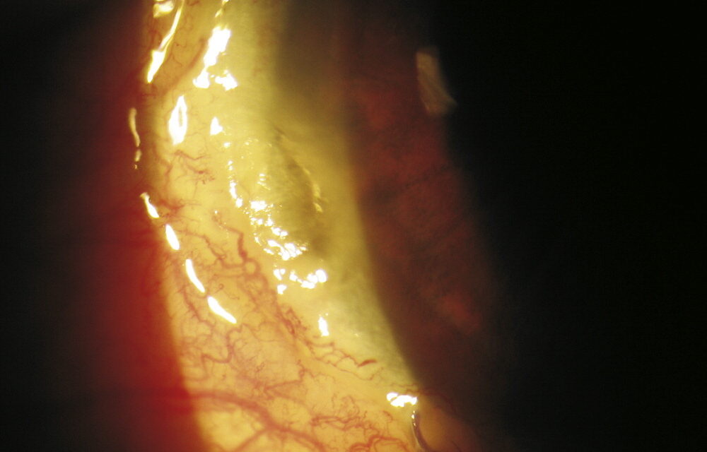
A corneal dellen (plural dellen) is a small, shallow, saucer-shaped depression at the corneal edge, right next to a raised area at the limbus.
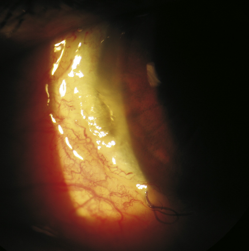
The corneal stroma beneath it is dehydrated and thin.
Patients report a foreign-body sensation or irritation, and the examiner sees a clear, glassy dimple that stains only slightly with fluorescein.
Most clear in a few days with simple treatment.
Why they form
The tear film needs a smooth surface to spread evenly.
When a raised lesion at the limbus interrupts the blink, tears are unable to wet the adjacent cornea, and the area dries out.
Epithelium remains intact, but the stroma loses water and thins.
The fluorescein dye pools in the depression, which shows up brightly, while the epithelium itself is not stained.
This distinguishes dellen from epithelial erosions and ulcers.
The name comes from the German word for a dent, and the term “Fuchs’ dimples” is also used.
Common settings
- Pterygium and pinguecula, the most frequent lesions (see pterygium)
- Filtering blebs after glaucoma surgery
- Subconjunctival hemorrhage and conjunctival chemosis (see chemosis)
- Postoperative swelling after strabismus or cataract surgery
- Limbal dermoids, tumors, and nodules
- Ocular surface disease such as dry eye, which makes dellen more likely
- Prolonged patching or lagophthalmos
All-fit Slit-Lamp Adapter
Record and share exactly what you see at the slit lamp. One adapter fits any slit lamp or surgical microscope — and any smartphone.
From Choroida — the team behind this siteWhat the examiner sees
- A well-demarcated, oval or crescent area of corneal thinning next to the lesion, with clear borders
- Intact epithelium, or slight punctate staining
- Fluorescein pooling in the depression
- Often no or little inflammation, and the anterior chamber is quiet
- Corneal thickness reduced, but rarely more than 50 percent
Differential diagnosis
Marginal keratitis and peripheral ulcerative keratitis are inflammatory, with infiltrates, epithelial defects and pain, and they must not be missed (see peripheral ulcerative keratitis).
Terrien marginal degeneration produces a gutter in the superior or inferior periphery with a lipid line, in older patients, and is non-inflammatory but progressive.
Dellen resolve quickly and do not progress.
Treatment
- Lubricate generously with preservative-free ointment or drops, so that the area stays wet. Frequent drops during the day with ointment at night often work in a matter of days.
- A pressure patch for a day or two helps hydrate the cornea in more resistant cases, but it should not be used where infection is possible.
- Treat the cause of the elevation, for example reducing bleb size or resolving chemosis, or, where it is the cause of recurrent dellen, excising a pterygium.
- Avoid steroids on an area of thinning unless there is a clear need.
How to examine a dellen
Use a narrow slit beam to see the depth of the depression, and compare the thickness of the cornea at the dellen with the adjacent area. Stain with fluorescein and watch the dye pool in the dip while the surrounding epithelium stays clear. Look at the lesion at the limbus that is causing the problem, and ask whether it has changed recently, for example after surgery or a hemorrhage. Check the tear film and lid position, since dry eye and incomplete blinking make dellen more likely.
Danger signs that point elsewhere
A dellen is painless or mildly irritating, quiet, and does not have an infiltrate. Pain, redness, discharge, an epithelial defect with an infiltrate, or progressive thinning mean that another diagnosis must be considered, including marginal keratitis, peripheral ulcerative keratitis, and infectious ulcer. Do not patch an eye if there is any suspicion of infection, and do not start steroids until the cause is clear.
Advice for the patient
Explain that the dimple forms because the tears cannot spread over the area, that it is not an infection, and that it should heal within days with frequent lubricants. Ask the patient to return if symptoms worsen, and to use the ointment at night until the cornea has recovered. If the cause is a lesion that will persist, such as a pterygium, discuss the options for removing it so that the problem does not recur.
Dellen in special situations
After strabismus surgery, a swollen conjunctiva near the limbus can produce a dellen within days, and generous lubrication from the first postoperative day prevents most cases. Beside a filtering bleb, dellen may be a reason to consider revising a bulky bleb if lubricants fail. In patients with a pterygium, recurrent dellen can be an indication for excision when other problems are also present. Contact lens wearers with a dellen should rest from wear until the cornea has recovered.
Prognosis
Dellen usually resolve fully within days.
A faint scar may remain.
Perforation is extremely rare, and almost always occurs when dellen are misdiagnosed and treated as an inflammatory process.


Document what you see
Two smartphone imaging tools built for everyday clinic use — one for the slit lamp, one for the fundus.
From Choroida — the team behind this siteReferences
- Krachmer JH, Mannis MJ, Holland EJ, eds. Cornea. 3rd ed. Philadelphia: Elsevier; 2011.
- Kanski JJ, Bowling B. Clinical Ophthalmology: A Systematic Approach. 7th ed. London: Elsevier; 2011.
- Yanoff M, Duker JS. Ophthalmology. 5th ed. Philadelphia: Elsevier; 2018.
- Albert DM, Miller JW, Azar DT, Blodi BA, eds. Albert and Jakobiec’s Principles and Practice of Ophthalmology. 3rd ed. Philadelphia: Saunders; 2008.
