
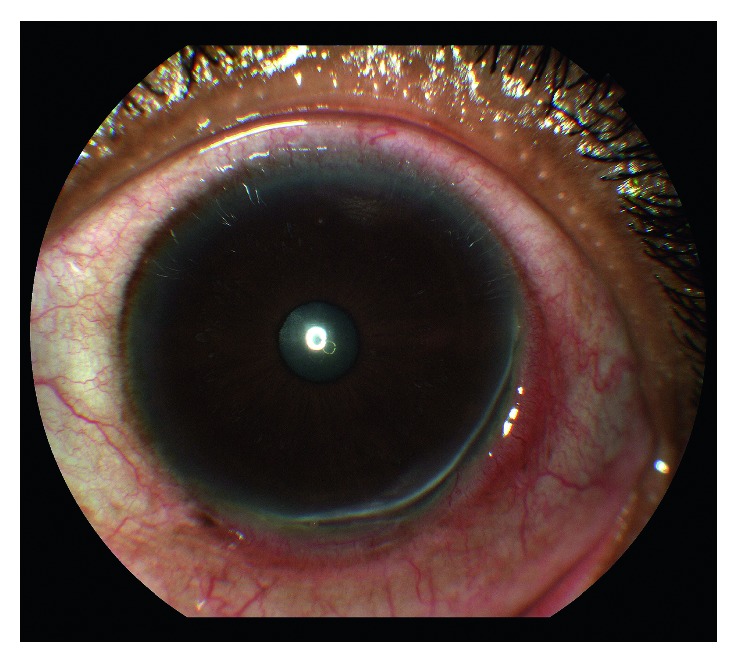
Peripheral ulcerative keratitis (PUK) is a crescent-shaped, progressive thinning and ulceration of the peripheral cornea.
It is one of the ophthalmic findings that most reliably signals serious, sometimes life-threatening, systemic autoimmune disease.
Roughly half of cases are associated with an underlying systemic connective tissue or vasculitic disorder, which is why PUK deserves a fundamentally different level of urgency and workup than an ordinary peripheral corneal thinning process.
Pathogenesis
The peripheral cornea sits adjacent to the limbal vasculature and is uniquely exposed to circulating immune complexes and inflammatory mediators in a way the avascular central cornea is not, which is part of why autoimmune and vasculitic disease so often manifests here first.
Immune complex deposition activates complement and recruits inflammatory cells that release collagenases and other proteolytic enzymes, progressively destroying the peripheral corneal stroma and producing the characteristic crescentic ulceration with an overhanging, undermined leading edge.
This same mechanism explains why PUK so often coexists with scleritis, since the adjacent sclera shares the same limbal vascular supply and is exposed to the same circulating immune complexes driving the corneal process.
Systemic Associations
- Rheumatoid arthritis — the most common systemic association overall, particularly in longstanding, seropositive, erosive disease
- Granulomatosis with polyangiitis (formerly Wegener’s) — PUK can be a presenting feature, and its appearance should prompt urgent ANCA testing and systemic workup
- Relapsing polychondritis — cartilage inflammation elsewhere (ears, nose, airway) alongside the ocular finding
- Systemic lupus erythematosus
- Polyarteritis nodosa and other systemic vasculitides
- Mooren’s ulcer — an idiopathic form without systemic disease, a diagnosis of exclusion once the above have been ruled out
PUK associated with active systemic vasculitis carries a serious prognosis if untreated, with reported mortality driven by the underlying systemic disease (cardiac and renal involvement) rather than the eye finding itself, which is the central reason PUK triggers an urgent, thorough systemic workup rather than purely local ocular treatment.
All-fit Slit-Lamp Adapter
Record and share exactly what you see at the slit lamp. One adapter fits any slit lamp or surgical microscope — and any smartphone.
From Choroida — the team behind this siteClinical Presentation
Patients present with pain, redness, photophobia, and tearing localized to the peripheral cornea, sometimes with visible thinning or a grayish infiltrate visible even without magnification in advanced cases.
Progression can be rapid: untreated PUK can lead to corneal perforation within days to weeks, when driven by an active systemic vasculitic process rather than the more indolent course sometimes seen in idiopathic Mooren’s ulcer.
Pain that seems disproportionate to the visible degree of corneal involvement, or that fails to improve with treatments aimed at more common causes of peripheral corneal irritation, should prompt reconsideration of the diagnosis and a more urgent, systemic-focused evaluation.
Exam Findings
- Crescent-shaped peripheral corneal ulceration with stromal thinning
- An undermined, overhanging central edge to the ulcer, a characteristic feature
- Adjacent limbal and conjunctival injection
- Absence of a clear infectious epithelial defect pattern (helping distinguish it from infectious keratitis)
- Associated scleritis in a meaningful proportion of cases, particularly when systemic vasculitis is the driver
Differential Diagnosis
- Terrien’s marginal degeneration — peripheral thinning but non-inflammatory, painless, slowly progressive, with an intact epithelium
- Infectious peripheral keratitis — positive culture, typically a clearer infectious trigger (contact lens wear, trauma)
- Marginal (staphylococcal hypersensitivity) keratitis — separated from the limbus by a clear zone, less destructive
- Pellucid marginal degeneration — inferior thinning with characteristic topography, non-inflammatory
- Neurotrophic keratopathy — reduced corneal sensation, different ulcer morphology
Diagnostic Evaluation
Any new case of PUK warrants a systemic workup, even in a patient without a known prior diagnosis of autoimmune disease, because PUK can be the presenting feature of a systemic vasculitis.
Complete blood count, inflammatory markers (ESR, CRP), rheumatoid factor, ANCA, antinuclear antibody, complement levels, and renal function testing are typically obtained, with the exact panel tailored to the clinical picture and guided by rheumatology input.
Mooren’s ulcer remains a diagnosis of exclusion, reached only once this systemic workup is negative.
Because a patient can present with PUK as their very first sign of an otherwise undiagnosed systemic vasculitis, urgent rheumatology involvement at the time of diagnosis — rather than after results return — allows the systemic workup and any necessary treatment to proceed in parallel rather than sequentially, which can matter when the underlying disease is progressing quickly.
Management
Systemic immunosuppression — often starting with corticosteroids and escalating to steroid-sparing agents (methotrexate, cyclophosphamide, or biologic agents depending on the underlying disease and severity) — is the cornerstone of treatment when PUK is associated with systemic vasculitis, coordinated closely with rheumatology, because controlling the systemic disease is what ultimately controls the ocular process.
Topical corticosteroids have a more limited and sometimes even counterproductive role, because they can worsen stromal melting in some cases without addressing the underlying immune complex-driven process.
Local measures — lubrication, bandage contact lenses, and tissue adhesive or patch grafting for impending or actual perforation — protect the ocular surface while systemic treatment takes effect.
Urgent surgical intervention (conjunctival resection, tectonic grafting) is reserved for eyes with perforation or severe, progressive thinning threatening globe integrity.
Because untreated systemic disease can also affect the fellow eye or recur even after an initial episode appears controlled, long-term rheumatologic follow-up continues well beyond resolution of the acute corneal findings, with any recurrence of ocular symptoms treated as a potential signal of renewed systemic disease activity.


Document what you see
Two smartphone imaging tools built for everyday clinic use — one for the slit lamp, one for the fundus.
From Choroida — the team behind this siteReferences
- Foster CS, Sainz de la Maza M. Peripheral ulcerative keratitis. In: The Sclera.
- Galor A, Thorne JE. Scleritis and peripheral ulcerative keratitis. Rheumatic Disease Clinics of North America.
- Tauber J, Sainz de la Maza M, Hoang-Xuan T, Foster CS. An analysis of therapeutic decision making regarding immunosuppressive chemotherapy for peripheral ulcerative keratitis. Cornea.
- American Academy of Ophthalmology. Basic and Clinical Science Course, Section 8: External Disease and Cornea.
Test yourself
A few questions straight from this article.
-
What is the characteristic corneal lesion of peripheral ulcerative keratitis?
Peripheral ulcerative keratitis is a crescent-shaped, progressive thinning and ulceration of the peripheral cornea. -
Roughly what proportion of peripheral ulcerative keratitis cases have an underlying systemic connective tissue or vasculitic disorder?
Roughly half of cases are associated with an underlying systemic connective tissue or vasculitic disorder, which is why the workup is urgent. -
Which mechanism drives stromal destruction in peripheral ulcerative keratitis?
Immune complex deposition activates complement and recruits cells that release collagenases and other proteolytic enzymes, destroying peripheral stroma. -
Which systemic disease is the most common association of peripheral ulcerative keratitis?
Rheumatoid arthritis is the most common systemic association overall, particularly in longstanding, seropositive, erosive disease. -
Which slit-lamp feature is characteristic of the ulcer in peripheral ulcerative keratitis?
An undermined, overhanging central edge to the crescentic ulcer is a characteristic feature of peripheral ulcerative keratitis. -
Which feature distinguishes Terrien's marginal degeneration from peripheral ulcerative keratitis?
Terrien's marginal degeneration causes peripheral thinning that is non-inflammatory, painless and slowly progressive, with an intact epithelium. -
Untreated peripheral ulcerative keratitis driven by active systemic vasculitis can perforate over which timeframe?
Progression can be rapid, with corneal perforation within days to weeks when an active systemic vasculitic process is driving the disease. -
What is the cornerstone of treatment for peripheral ulcerative keratitis associated with systemic vasculitis?
Systemic immunosuppression, often corticosteroids escalating to steroid-sparing or biologic agents, controls the systemic disease and so the cornea. -
Why is the role of topical corticosteroids limited in peripheral ulcerative keratitis?
Topical steroids can worsen stromal melting in some cases and do not address the underlying immune complex-driven process. -
In peripheral ulcerative keratitis with active systemic vasculitis, reported mortality is driven mainly by what?
Mortality reflects the underlying systemic disease, with cardiac and renal involvement rather than the corneal finding itself.

Writes and reviews the clinical articles on Educate. Every post is checked against peer-reviewed sources before it goes live.


