
A Soemmering ring is a circular, doughnut-shaped opacity at the periphery of the capsular bag.
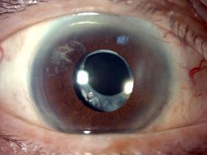
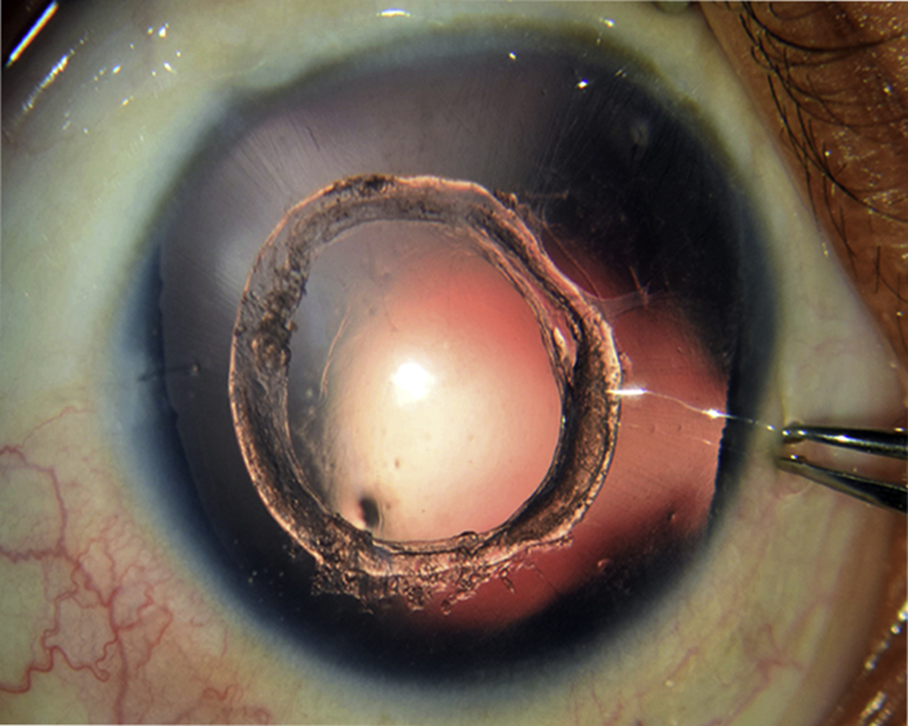
It forms after the lens contents have been removed from the eye but the capsular bag has been left in place, and it is composed of residual cortex and proliferating lens epithelial cells trapped between the anterior and posterior capsule remnants.
How it forms
In extracapsular surgery and phacoemulsification, the surgeon removes the nucleus and cortex through a hole in the anterior capsule, but some lens epithelial cells always remain.
They sit at the equator, where the anterior and posterior capsule meet, and in the weeks after surgery they proliferate.
The cells lay down new lens fibers and produce a ring of soft, translucent, white material.
The ring is largest when the anterior capsule opening is small and the bag is closed, trapping the cells inside.
The ring was described in the early nineteenth century by the German anatomist Samuel Thomas von Soemmerring, who studied eyes after couching and early extraction.
How it looks
At the slit lamp, with the pupil dilated, the ring appears as a white or grayish band of material around the periphery of the bag.
It may be seen behind the iris, as a thick circular mass that sits outside the optic of the intraocular lens.
In a child after lensectomy, the ring can grow into the visual axis.
Soemmering rings are usually asymptomatic.
They do not cause a visual problem unless the material migrates, or the bag becomes opacified.
All-fit Slit-Lamp Adapter
Record and share exactly what you see at the slit lamp. One adapter fits any slit lamp or surgical microscope — and any smartphone.
From Choroida — the team behind this siteWhat it can lead to
- Posterior capsule opacification, since the same cells that make up the ring migrate toward the center of the posterior capsule (see Elschnig pearls and posterior capsule opacification)
- Intraocular lens decentration or tilt, particularly when the ring is large and asymmetrical and pushes the IOL optic off center
- Pseudo-ring appearance in the red reflex, with glare and halos in some patients
- In pediatric surgery, visual-axis opacification that needs additional surgery
Related situations
A similar ring can form after trauma that ruptures the lens capsule, when lens matter is resorbed leaving a ring of capsule and cortex, and after spontaneous resorption of a hypermature cataract.
It can also be seen in some children with congenital cataract that underwent partial removal.
Do you need to treat it?
Most Soemmering rings need no treatment.
Intervention is considered when the ring causes symptoms or complications.
- Nd:YAG capsulotomy for associated posterior capsule opacification (see YAG laser capsulotomy)
- Surgical removal in children, with posterior capsulotomy and anterior vitrectomy when the visual axis is involved
- Repositioning or exchanging a decentered IOL if it causes visual disturbance
How to reduce the chance of a ring
Careful cortical clean-up and polishing of the capsule at the end of the operation, a well-centered continuous curvilinear capsulorhexis that overlaps the edge of the IOL optic, and the use of IOLs with a square optic edge all reduce the risk of cell migration and capsular opacification.
How to examine for a ring
Dilate the pupil fully and examine at the slit lamp with direct illumination and retroillumination. The ring lies behind the iris, so it is invisible in an undilated eye. Look for a doughnut-shaped band of whitish, sometimes pearly, material at the periphery of the bag, outside the edge of the lens optic. In a child, check whether the ring extends toward the visual axis and whether the posterior capsule behind the optic remains clear. Anterior segment OCT can show the thickness of the material and its relation to the lens haptics when the view is poor.
Pitfalls
- Do not mistake a ring for a hypermature cataract or a dislocated lens
- Do not treat a clear, asymptomatic ring with laser, because capsulotomy is not needed unless the central capsule is opaque
- Do not forget that a ring is a reservoir of lens epithelial cells, so late posterior capsule opacification is more likely when it is large
Explain to the patient that the ring is a normal healing response and that the planned follow-up will detect any change.
Ring after trauma or in children
After a penetrating injury that ruptures the lens capsule, the cortex can swell, absorb, and leave a ring that persists for life. In children, the ring often grows faster than in adults, and it can extend into the pupillary area, causing amblyopia if the visual axis becomes opaque. Close follow-up with regular dilated examinations is needed after pediatric cataract surgery, and parents should be told that a second small operation to clear the axis may be required.
Prognosis
The ring itself is benign.
Most patients will not know that they have one, and the visual outcome after surgery is excellent in the vast majority of cases.


Document what you see
Two smartphone imaging tools built for everyday clinic use — one for the slit lamp, one for the fundus.
From Choroida — the team behind this siteReferences
- Apple DJ, Solomon KD, Tetz MR, et al. Posterior capsule opacification. Surv Ophthalmol. 1992;37:73-116.
- Wormstone IM, Wang L, Liu CS. Posterior capsule opacification. Exp Eye Res. 2009;88:257-269.
- Nishi O. Posterior capsule opacification. Part 1: experimental investigations. J Cataract Refract Surg. 1999;25:106-117.
- Kappelhof JP, Vrensen GF. The pathology of after-cataract. Acta Ophthalmol Suppl. 1992;205:13-24.
