
Type 2 macular telangiectasia, often called MacTel type 2, has undergone a significant conceptual shift in how it is understood: once viewed primarily as a vascular retinal condition given its telangiectatic name, growing evidence now points toward an underlying neurodegenerative process, specifically involving Müller cell dysfunction, as the more fundamental driver, with the visible vascular changes representing a secondary consequence rather than the primary problem.
A Bilateral, Slowly Progressive Condition
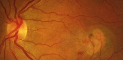
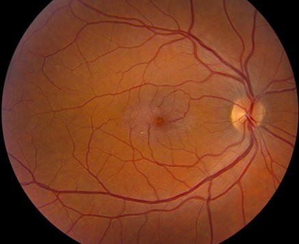
This condition typically presents in middle-aged to older adults, is usually bilateral, though sometimes asymmetric, and follows a slowly progressive course over years, affecting the parafoveal retina in a characteristic pattern distinct from more common macular conditions such as age-related macular degeneration or diabetic macular edema.
Clinical Features
- Subtle grayish discoloration of the parafoveal retina, often temporal to the fovea, representing one of the earliest and most characteristic findings, though easily overlooked without specific familiarity with this appearance
- Crystalline deposits can be seen scattered within the affected parafoveal retina in some cases
- Right-angle venules, retinal venules making an abrupt, distinctive right-angle turn as they course toward the fovea, represent a characteristic, specifically recognized vascular sign of this condition
- Telangiectatic retinal vessels within the affected area, the feature the condition is named for, though as understanding of the disease has evolved, these vascular changes are increasingly viewed as a downstream consequence of underlying retinal tissue dysfunction rather than the primary driver of disease
- Gradual, progressive central vision loss develops over the disease course, related to progressive photoreceptor and retinal pigment epithelial changes within the affected parafoveal zone
Fundus Explorer Pro
Photograph the retinal findings described here with the phone already in your pocket — 22 D optics and built-in illumination in one handheld unit.
From Choroida — the team behind this siteThe Neurodegenerative Reconceptualization
Growing evidence, including findings from optical coherence tomography showing progressive loss of the ellipsoid zone and outer retinal structure preceding more obvious vascular change, along with other supporting research, has shifted understanding of this condition toward a primary retinal neurodegenerative process, with Müller cell dysfunction implicated as a central, early mechanism, and the visible telangiectatic vascular changes understood as accompanying rather than causative of the broader retinal tissue dysfunction.
This reconceptualization matters because it has shaped the direction of research into potential future treatments, moving attention beyond purely vascular-targeted therapy toward approaches that might address the underlying neurodegenerative process more directly.
Complications
A subset of patients develops neovascularization as a complication of the underlying disease process, sometimes referred to as the proliferative stage of the condition, which can produce more acute vision loss related to exudation or hemorrhage, distinct from the more typical slow, gradual vision decline the non-neovascular disease course produces.
Diagnostic Evaluation
- Dilated fundus examination, looking specifically for the characteristic subtle parafoveal grayish discoloration and, when present, crystalline deposits and right-angle venules
- Optical coherence tomography, demonstrating characteristic parafoveal cavitation and, in more advanced cases, ellipsoid zone disruption, supporting diagnosis and monitoring of disease progression
- Fluorescein angiography can demonstrate the characteristic telangiectatic vessels and, when present, any associated neovascularization
- Optical coherence tomography angiography, where available, provides additional, detailed characterization of the vascular changes without requiring dye injection
Management
Non-Neovascular Disease
No proven treatment currently halts the progressive, non-neovascular course of this condition, and management for this stage centers on monitoring disease progression and providing appropriate low vision support and counseling as visual function gradually declines over time.
Managing Neovascular Complications
When neovascularization develops as a complication, anti-VEGF therapy is used, following similar principles to its use for neovascular complications in other retinal conditions, aiming to control the acute exudative or hemorrhagic component this complication produces.
What the OCT shows
OCT is the key test. In the early stages, there are small hyporeflective cavities in the inner and outer retina near the fovea, without a lot of thickening. Later, the ellipsoid zone breaks up, and the outer retina thins. In some cases, a pigmented plaque forms, and neovascularization may develop. Autofluorescence shows increased signal in the macula, and blue-light reflectance is reduced, which gives a characteristic appearance.
Counselling
Tell patients that the disease is slowly progressive, that it usually affects both eyes, and that there is currently no proven treatment for the non-neovascular form. An encapsulated cell therapy implant has been studied in clinical trials. Encourage participation in research registries, provide low vision support, and use anti-VEGF for neovascular disease.
Prognosis
Type 2 macular telangiectasia follows a slowly progressive course over years to decades, with gradual central vision decline in most affected patients, though the rate and ultimate severity of vision loss vary considerably between individuals.
Ongoing research into the underlying neurodegenerative mechanism driving this condition continues to shape the search for more effective future treatments, reflecting the field’s evolving understanding of this condition as fundamentally a retinal neurodegenerative disease rather than a primarily vascular one.


Document what you see
Two smartphone imaging tools built for everyday clinic use — one for the slit lamp, one for the fundus.
From Choroida — the team behind this siteReferences
- Yannuzzi LA, Bardal AM, Freund KB, Chen KJ, Eandi CM, Blodi B. Idiopathic macular telangiectasia. Arch Ophthalmol. 2006;124:450-460.
- Powner MB, Gillies MC, Tretiach M, et al. Perifoveal Muller cell depletion in a case of macular telangiectasia type 2. Ophthalmology. 2010;117:2407-2416.
- Charbel Issa P, Gillies MC, Chew EY, et al. Macular telangiectasia type 2. Prog Retin Eye Res. 2013;34:49-77.
- Clemons TE, Gillies MC, Chew EY, et al. Medical characteristics of patients with macular telangiectasia type 2 (MacTel Type 2) MacTel Project Report No. 3. Ophthalmic Epidemiol. 2013;20:109-113.
