
Fundus flavimaculatus and Stargardt disease are now understood to represent points along a single genetic and clinical spectrum rather than two separate diseases, and appreciating this relationship, along with the specific fundus features that distinguish the more diffuse, later-onset flavimaculatus pattern from classic macula-predominant Stargardt disease, shapes both prognosis and genetic counseling for affected families.
A Shared Genetic Basis
Both conditions result from mutations in the ABCA4 gene, inherited in an autosomal recessive pattern, which encodes a protein important for normal photoreceptor outer segment metabolism, and dysfunction of this protein leads to accumulation of a toxic byproduct, lipofuscin, within the retinal pigment epithelium, producing the characteristic yellow-white flecks and progressive retinal pigment epithelial and photoreceptor degeneration seen in both presentations.
Classic Stargardt Disease
- Typically presents earlier, often in the first or second decade of life, with progressive central visual loss
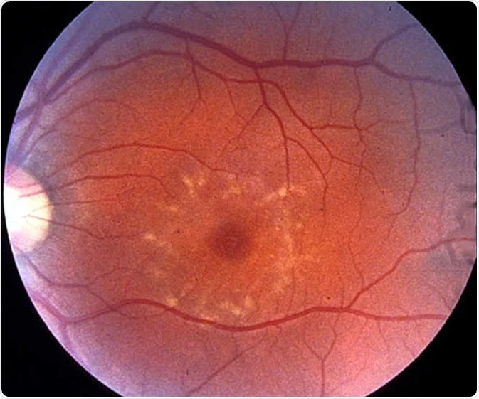
- Yellow-white, pisciform (fish-tail shaped) flecks concentrated around the macula, with the macula itself often showing an atrophic, “beaten bronze” appearance in more advanced disease
- Central visual acuity loss is often the dominant, earliest, and most functionally significant symptom, given the macula-predominant distribution of disease
Fundus Explorer Pro
Photograph the retinal findings described here with the phone already in your pocket — 22 D optics and built-in illumination in one handheld unit.
From Choroida — the team behind this siteFundus Flavimaculatus
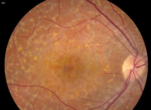
- Generally presents somewhat later, and the flecks are more diffusely distributed throughout the posterior pole and mid-peripheral retina, rather than being concentrated predominantly around the macula
- Central visual acuity can be relatively preserved for longer in some patients, particularly earlier in the disease course, compared with classic Stargardt disease, given the more diffuse rather than macula-concentrated pattern of involvement
- Represents, in current understanding, the same underlying disease process and gene as Stargardt disease, differing mainly in the distribution and timing of flecks and degeneration rather than reflecting a fundamentally distinct condition
The Dark Choroid Sign
A characteristic finding on fluorescein angiography in this disease spectrum is the “dark choroid” or “silent choroid” sign, in which the background choroidal fluorescence is abnormally suppressed, thought to result from lipofuscin accumulation within the retinal pigment epithelium blocking the normal transmission of choroidal fluorescence, and this sign, when present, is considered a fairly specific supportive finding for this disease spectrum.
Diagnostic Evaluation
- Dilated fundus examination characterizing the distribution, extent, and appearance of flecks and any macular atrophy
- Fundus autofluorescence imaging is particularly valuable, since the lipofuscin-laden flecks and areas of retinal pigment epithelial atrophy show characteristic, distinctive patterns of increased or decreased autofluorescence, often more sensitive for detecting and mapping disease extent than color fundus photography alone
- Optical coherence tomography demonstrates outer retinal and retinal pigment epithelial changes and can help stage disease severity and track progression over time
- Electroretinography helps assess the extent of generalized retinal (rod and cone) dysfunction, which correlates with prognosis and can help distinguish more macula-limited from more generalized disease patterns
- Genetic testing for ABCA4 mutations confirms the diagnosis and supports genetic counseling for affected families
Prognosis and Disease Course
Prognosis in this disease spectrum is quite variable, correlating with the age of onset (earlier onset generally associated with more severe, rapidly progressive disease), the extent of flecks and atrophy at presentation, and results of electroretinography, with more generalized rod-cone dysfunction on electroretinography associated with a worse overall visual prognosis than disease that remains predominantly confined to the macula.
Both classic Stargardt disease and fundus flavimaculatus ultimately tend toward progressive visual decline over years to decades, though the rate and pattern of progression vary considerably between individuals, even within the same family carrying the same underlying mutation.
Management
There is currently no treatment that reverses or halts the underlying genetic and metabolic disease process, so management is centered on supportive care and monitoring.
- Low vision rehabilitation services, becoming increasingly important as central vision declines
- Genetic counseling for affected families, given the autosomal recessive inheritance pattern and implications for future children and other family members
- Avoidance of high-dose vitamin A supplementation has been specifically recommended in this disease spectrum, based on concern that vitamin A may contribute to further lipofuscin accumulation, an important and specific counseling point that differs from general retinal health advice
- Ongoing monitoring of disease progression, both to track the individual patient’s course and given the active research interest in emerging gene-specific and other therapeutic approaches for this genetically well-characterized disease
Clinical Significance
Understanding fundus flavimaculatus and Stargardt disease as points on a single spectrum, rather than as entirely separate diagnoses, supports more accurate genetic counseling and prognosis discussion, and reinforces the value of fundus autofluorescence and electroretinography, alongside standard fundus examination, in fully characterizing where along this spectrum a given patient’s disease falls.


Document what you see
Two smartphone imaging tools built for everyday clinic use — one for the slit lamp, one for the fundus.
From Choroida — the team behind this siteReferences
- Tanna P, Strauss RW, Fujinami K, Michaelides M. Stargardt disease: clinical features, molecular genetics, animal models and therapeutic options. Br J Ophthalmol. 2017;101:25-30.
- Fishman GA, Farbman JS, Alexander KR. Delayed rod dark adaptation in patients with Stargardt’s disease. Ophthalmology. 1991;98:957-962.
- Lois N, Holder GE, Bunce C, Fitzke FW, Bird AC. Phenotypic subtypes of Stargardt macular dystrophy-fundus flavimaculatus. Arch Ophthalmol. 2001;119:359-369.
- Fishman GA, Stone EM, Grover S, Derlacki DJ, Haines HL, Hockey RR. Variation of clinical expression in patients with Stargardt dystrophy and sequence variations in the ABCA4 gene. Arch Ophthalmol. 1999;117:504-510.
