
Angle recession glaucoma is a genuinely important reason blunt ocular trauma, even trauma that seemed to heal without any obvious lasting problem, deserves lifelong follow-up, since the glaucoma it causes can develop silently and present for the first time years, even decades, after the original injury has been forgotten.
What Happens at the Time of Injury
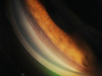
Blunt trauma to the eye can cause a tear between the longitudinal and circular fibers of the ciliary body, a structural injury to the anterior chamber angle called angle recession, which widens the ciliary body band as seen on gonioscopy compared with the fellow, uninjured eye.
This structural change itself does not necessarily cause immediate pressure elevation, but the same traumatic event that caused the recession often also damages the adjacent trabecular meshwork, and it is this trabecular meshwork scarring and dysfunction, developing and progressing gradually over subsequent years, that eventually produces glaucoma in a subset of affected eyes.
Why the Glaucoma Can Be So Delayed
The gap between the traumatic injury and the eventual development of elevated intraocular pressure can be substantial, sometimes many years or even decades, which is why a patient with angle recession glaucoma may not connect their new glaucoma diagnosis with an old injury from childhood or early adulthood unless specifically asked, and why gonioscopic examination, comparing the two eyes, is what reveals the connection.
All-fit Slit-Lamp Adapter
Record and share exactly what you see at the slit lamp. One adapter fits any slit lamp or surgical microscope — and any smartphone.
From Choroida — the team behind this siteCommon Mechanisms of Injury
- Sports-related trauma, including projectiles such as a ball, a common and frequently cited mechanism
- Airbag deployment during a motor vehicle accident
- Fist or other blunt-object assault
- Any other blunt impact directly to the eye or orbit
Gonioscopic Findings
- A widened ciliary body band in the area of recession, visible on careful gonioscopy, typically most pronounced in one sector corresponding to the area of direct impact rather than uniformly around the entire angle circumference
- Comparison with the fellow, uninjured eye is essential, since some degree of angle width variation exists normally, and it is the asymmetry between the two eyes of the same patient that confirms a traumatic, rather than simply anatomic, origin
- Other signs of prior blunt trauma may be present concurrently, including iris sphincter tears, phacodonesis from zonular injury, or a Vossius ring (a pigment ring on the anterior lens capsule from iris pigment epithelium transfer at the moment of impact)
Clinical Course
Glaucoma develops in only a subset of eyes with angle recession, and the risk correlates to some degree with the extent of recession, with more extensive recession (involving a greater circumferential portion of the angle) generally carrying higher long-term glaucoma risk.
Once established, angle recession glaucoma tends to behave similarly to primary open-angle glaucoma in its response to treatment, though the underlying trabecular meshwork damage means it can sometimes be more resistant to typical medical therapy in some patients.
Diagnosis and Monitoring
- A history of any significant blunt ocular trauma, however remote, should prompt gonioscopic comparison of both angles at baseline and periodic reassessment of intraocular pressure over the following years and decades
- Any patient found to have gonioscopic angle recession, regardless of current intraocular pressure, warrants lifelong periodic monitoring for glaucoma, given the potential for very delayed onset
- Optic nerve assessment and visual field testing are incorporated into monitoring once any pressure elevation or optic nerve change is detected, following standard glaucoma evaluation principles
Management
Management of established angle recession glaucoma follows standard glaucoma treatment principles, beginning with topical aqueous-suppressant medical therapy, and progressing to laser or incisional surgery when medical therapy is inadequate to control pressure and protect the optic nerve.
Given the underlying trabecular meshwork scarring, selective laser trabeculoplasty may be somewhat less effective than in eyes without traumatic angle injury, an important consideration when selecting among treatment options for a given patient.
Prognosis
With lifelong awareness of the injury and appropriately sustained gonioscopic and pressure monitoring, angle recession glaucoma can generally be detected before significant optic nerve damage occurs, allowing timely treatment.
The greatest risk lies in patients whose traumatic history is not elicited or whose angle recession is never identified, allowing glaucoma to progress undetected over years, which is why asking about any history of significant eye trauma, however long ago, remains a routine and valuable part of comprehensive eye examination.


Document what you see
Two smartphone imaging tools built for everyday clinic use — one for the slit lamp, one for the fundus.
From Choroida — the team behind this siteReferences
- Tesluk GC, Spaeth GL. The occurrence of glaucoma after blunt trauma: a retrospective and prospective study. Ophthalmic Surg. 1985;16:279-285.
- Sihota R, Sood NN, Agarwal HC. Traumatic glaucoma. Acta Ophthalmol Scand. 1995;73:252-254.
- Kaufman JH, Tolpin DW. Glaucoma after traumatic angle recession: a ten-year prospective study. Am J Ophthalmol. 1974;78:648-654.
- Girkin CA, McGwin G Jr, Long C, Morris R, Kuhn F. Glaucoma after ocular contusion: a cohort study of the United States Eye Injury Registry. J Glaucoma. 2005;14:470-473.
