
Filamentary keratitis produces strands of degenerated epithelial cells and mucus that adhere to the corneal surface at one end and trail free at the other, moving with each blink and pulling on the underlying corneal epithelium in a way that causes disproportionately severe foreign body sensation for what can be, on first glance, a subtle-looking finding.
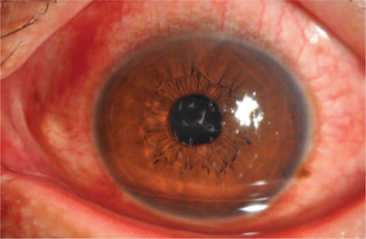
How Filaments Form
Filaments develop when areas of unstable or devitalized corneal epithelium, combined with an abnormal, often deficient or excessively viscous, mucus layer of the tear film, allow strands of epithelial cells and mucin to twist together and adhere at one point to the corneal surface.
The filament’s free end moves with blinking, and its attached end tugs on the underlying epithelium with each movement, which is why filamentary keratitis causes a degree of foreign body sensation and pain that seems disproportionate to the size of the finding on examination.
Causes and Associations
- Severe dry eye disease, including aqueous-deficient dry eye and Sjögren syndrome, among the most common underlying causes
- Prolonged patching or eye closure, including after surgery
- Superior limbic keratoconjunctivitis
- Neurotrophic keratitis, given the combination of poor tear film and reduced epithelial turnover
- Prolonged contact lens wear
- Recurrent corneal erosion
- Cranial nerve palsies affecting blink or tear function
All-fit Slit-Lamp Adapter
Record and share exactly what you see at the slit lamp. One adapter fits any slit lamp or surgical microscope — and any smartphone.
From Choroida — the team behind this siteClinical Features
- Foreign body sensation, tearing, photophobia, and blepharospasm, often out of proportion to the visible clinical findings
- Fine, comma-shaped or thread-like strands adhering to the corneal surface, best seen with fluorescein or rose bengal staining, which the filaments characteristically take up
- Symptoms typically worsen with blinking, as the filament’s free end catches and pulls
- Often associated with visible signs of the underlying dry eye or surface disease, such as reduced tear breakup time and punctate epithelial staining elsewhere on the cornea
Diagnosis
Diagnosis is made on slit-lamp examination, with fluorescein or rose bengal staining highlighting the filaments clearly against the corneal surface.
A workup for the underlying cause, particularly severe dry eye disease, is important, since treating filaments without addressing the underlying tear film abnormality leads to recurrence.
Management
Mechanical Removal
Gentle mechanical debridement of the filaments, using fine forceps at the slit lamp after topical anesthesia, provides immediate symptomatic relief, though filaments can and often do recur if the underlying cause is not addressed, sometimes within days.
Lubrication and Mucolytics
- Preservative-free artificial tears and lubricating ointment, used frequently, address the underlying tear film instability
- Topical mucolytic agents, such as acetylcysteine, can help dissolve the mucous component of filaments and reduce their formation, and are used both for acute treatment and as ongoing prophylaxis in recurrent cases
Bandage Contact Lens
A bandage contact lens can provide mechanical protection and comfort in more severe or recurrent cases, reducing the mechanical irritation from blinking over an unstable epithelial surface while the underlying dry eye is treated.
Treating the Underlying Cause
Definitive management addresses the cause of the abnormal tear film and epithelium: aggressive dry eye treatment, including punctal occlusion and anti-inflammatory therapy for significant aqueous-deficient dry eye, management of any eyelid or lid closure abnormality, and treatment of an underlying neurotrophic component if present.
Prognosis
Filamentary keratitis responds well to treatment in most cases, with rapid symptomatic relief after debridement, though the underlying condition, often chronic dry eye disease, requires ongoing management to prevent recurrence.
Patients with severe, poorly controlled underlying disease, such as advanced Sjögren syndrome, may have a more chronic, relapsing course requiring sustained, multi-pronged ocular surface treatment.


Document what you see
Two smartphone imaging tools built for everyday clinic use — one for the slit lamp, one for the fundus.
From Choroida — the team behind this siteReferences
- Zaidman GW, Geeraets R, Paylor RR, Ferry AP. The histopathology of filamentary keratitis. Arch Ophthalmol. 1985;103:1178-1181.
- Albietz JM, Lenton LM. Management of filamentary keratitis associated with aqueous-deficient dry eye. Optom Vis Sci. 2007;84:E96-E105.
- Wright P. Filamentary keratitis. Trans Ophthalmol Soc U K. 1975;95:260-262.
- Bron AJ, de Paiva CS, Chauhan SK, et al. TFOS DEWS II pathophysiology report. Ocul Surf. 2017;15:438-510.
