
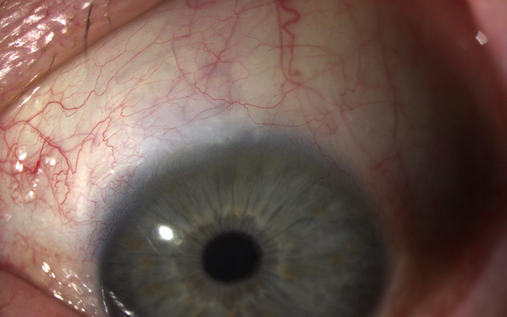
Canaloplasty is a non-penetrating glaucoma procedure that mechanically dilates and stents open Schlemm’s canal, restoring the eye’s own natural outflow pathway rather than creating a new drainage route the way trabeculectomy or tube shunt surgery, both discussed in their own dedicated articles on this site, do.
Because it does not enter the anterior chamber or create a filtering bleb, canaloplasty avoids many of the bleb-related complications, including infection risk and chronic bleb management, that make trabeculectomy such a demanding procedure to follow long-term.
Understanding where canaloplasty sits between medical therapy, MIGS, and traditional filtering surgery, discussed in its own dedicated article on this site, helps clarify why it has found a specific, if comparatively narrow, place in the modern glaucoma surgical toolkit.
The technique demands considerable surgical skill to thread the microcatheter successfully around the full circumference of a structure as small and delicate as Schlemm’s canal, which is part of why it has not been adopted as widely as some of the newer, technically simpler MIGS options.
Technique
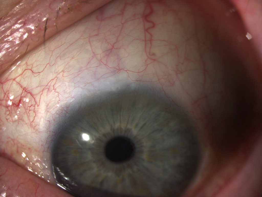
The surgeon dissects a deep scleral flap to expose Schlemm’s canal, then threads a flexible microcatheter with an illuminated tip circumferentially around the entire canal, visualizing its progress through the sclera as it advances.
A suture is attached to the microcatheter and drawn through the full circumference of the canal as the catheter is withdrawn, and this suture is then tensioned and tied to provide sustained circumferential dilation of the canal and the adjacent trabecular meshwork.
Because the procedure works entirely within the canal itself and does not breach the anterior chamber, it is classified as non-penetrating, in contrast to trabeculectomy’s deliberate, full-thickness opening into the anterior chamber.
Deep sclerectomy is a related non-penetrating technique that similarly avoids full-thickness anterior chamber entry, though without the circumferential catheterization and suture tensioning that specifically defines canaloplasty.
Mechanism of Pressure Reduction
Dilating Schlemm’s canal is thought to reduce resistance to aqueous outflow at the level of the juxtacanalicular trabecular meshwork, the tissue layer generally considered to contribute the most resistance to normal outflow in open-angle glaucoma.
The tensioned suture maintains this dilation over the long term, providing a sustained mechanical effect distinct from the more biological, cellular mechanism behind procedures like selective laser trabeculoplasty, discussed in its own dedicated article on this site.
Unlike trabeculectomy, canaloplasty does not rely on the formation and maintenance of a filtering bleb, which is the central reason it avoids the bleb-specific complications, including blebitis, discussed in its own dedicated article on this site, that represent a lifelong risk after successful trabeculectomy.
All-fit Slit-Lamp Adapter
Record and share exactly what you see at the slit lamp. One adapter fits any slit lamp or surgical microscope — and any smartphone.
From Choroida — the team behind this siteIndications and Patient Selection
- Open-angle glaucoma requiring surgical intervention, particularly in patients and surgeons prioritizing avoidance of a filtering bleb and its associated long-term risks and lifestyle restrictions
- Moderate rather than very advanced glaucoma, since canaloplasty’s pressure-lowering ceiling is generally lower than trabeculectomy’s, similar in this respect to the MIGS category discussed in its own dedicated article on this site
- Patients wishing to avoid or delay a bleb-dependent procedure for lifestyle reasons, including those who swim regularly or have occupational or personal concerns about long-term bleb maintenance
Advanced glaucoma requiring a very low target pressure is generally better served by trabeculectomy or a tube shunt, mirroring the same general pressure-ceiling limitation discussed for MIGS procedures more broadly.
Complications
Hyphema is relatively common in the early postoperative period, reflecting blood reflux through the newly dilated, previously collapsed outflow channels as normal physiologic pressure gradients reassert themselves.
Descemet membrane detachment, discussed in its own dedicated article on this site, and suture-related complications, including suture breakage or cheese-wiring through the canal tissue, are recognized technique-specific risks.
The overall complication profile is generally considered milder than trabeculectomy’s, without the ongoing infection and bleb-leak risks that persist indefinitely after a successful filtering procedure, which is a central part of canaloplasty’s appeal despite its more limited pressure-lowering ceiling.
Outcomes
Canaloplasty achieves meaningful pressure reduction in appropriately selected eyes, generally more modest than trabeculectomy but without the same bleb-dependent long-term risk profile.
Long-term data, while growing, remains less extensive than the many decades of experience behind trabeculectomy, and canaloplasty is used by a smaller subset of glaucoma surgeons who have specifically trained in and adopted the technique, reflecting its greater technical demands compared to more widely practiced alternatives.
Patient counseling should reflect this trade-off honestly: a milder complication profile and no bleb to manage, weighed against a more modest pressure-lowering ceiling and a procedure available at fewer centers than trabeculectomy.


Document what you see
Two smartphone imaging tools built for everyday clinic use — one for the slit lamp, one for the fundus.
From Choroida — the team behind this siteReferences
- Lewis RA, von Wolff K, Tetz M, et al. Canaloplasty: circumferential viscodilation and tensioning of Schlemm’s canal using a flexible microcatheter for the treatment of open-angle glaucoma in adults. Journal of Cataract and Refractive Surgery.
- American Academy of Ophthalmology. Basic and Clinical Science Course, Section 10: Glaucoma.
- Brusini P. Canaloplasty in open-angle glaucoma surgery: a four-year follow-up. The Scientific World Journal.
Test yourself
A few questions straight from this article.
-
What does canaloplasty do to lower intraocular pressure in open-angle glaucoma?
Canaloplasty is a non-penetrating procedure that dilates and stents Schlemm's canal open, restoring the eye's own natural outflow pathway instead of creating a new drainage route. -
Which instrument is threaded circumferentially around Schlemm's canal during canaloplasty?
After dissecting a deep scleral flap, the surgeon threads a flexible microcatheter with an illuminated tip around the whole canal, tracking its progress through the sclera. -
Why is canaloplasty classified as a non-penetrating glaucoma procedure?
Canaloplasty operates entirely within Schlemm's canal and never breaches the anterior chamber, unlike trabeculectomy's deliberate full-thickness opening into it. -
Dilating Schlemm's canal is thought to reduce outflow resistance mainly at which tissue?
The juxtacanalicular trabecular meshwork is generally considered the tissue contributing most resistance to outflow in open-angle glaucoma, and canaloplasty reduces resistance at that level. -
Which feature specifically defines canaloplasty compared with deep sclerectomy?
Deep sclerectomy is a related non-penetrating technique, but the circumferential catheterization of the canal and the tensioned suture are what specifically define canaloplasty. -
Which glaucoma patient is generally better served by trabeculectomy or a tube shunt than by canaloplasty?
Canaloplasty's pressure-lowering ceiling is generally lower than trabeculectomy's, so advanced disease requiring a very low target pressure is better served by trabeculectomy or a tube shunt. -
What explains the hyphema commonly seen in the early period after canaloplasty?
Hyphema is relatively common early after canaloplasty and reflects blood refluxing through outflow channels that were previously collapsed, as normal physiologic pressure gradients reassert themselves. -
Which suture-related problem is a recognized technique-specific risk of canaloplasty?
Suture breakage and cheese-wiring through the canal tissue are recognized technique-specific complications of canaloplasty, alongside Descemet membrane detachment. -
How does canaloplasty's pressure-lowering mechanism differ from that of selective laser trabeculoplasty?
The tensioned suture maintains canal dilation long term, a sustained mechanical effect that differs from the more biological, cellular mechanism underlying selective laser trabeculoplasty. -
Which trade-off should patient counselling before canaloplasty honestly reflect?
Canaloplasty offers a milder complication profile and no bleb to manage, weighed against a more modest pressure-lowering ceiling and availability at fewer centres than trabeculectomy.
