
Tubulointerstitial nephritis and uveitis (TINU) syndrome combines acute tubulointerstitial nephritis with bilateral anterior uveitis, usually in adolescents and young women.
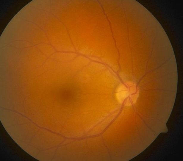
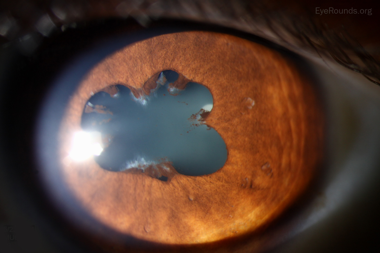
The kidney disease often comes first and can be mild enough to pass unnoticed, so the uveitis is sometimes what brings the diagnosis to light.
Ophthalmologists who recognize the pattern can save the patient a delayed nephrology referral.
What Is TINU Syndrome?
TINU was first described in 1975, and it remains an uncommon but probably underdiagnosed cause of uveitis in young patients.
The nephritis is usually acute and interstitial, with normal glomeruli on biopsy.
Systemic symptoms are often nonspecific: fatigue, weight loss, fever, and flank or abdominal pain.
Some patients have no renal symptoms at all, and the kidney involvement is found only on urinalysis or blood testing done because of the eye disease.
Ocular Features
Uveitis develops before, during, or after the nephritis, and the interval between the two can be several months in either direction.
- Bilateral anterior uveitis in most cases, though it can start unilaterally
- Nongranulomatous inflammation with small keratic precipitates
- Anterior chamber cells and flare, sometimes with a hypopyon in severe cases
- Posterior involvement is uncommon but has been reported, including vitritis and disc edema
- Recurrence is frequent, and some patients have a chronic relapsing course
The uveitis usually responds to topical corticosteroids, but recurrent or bilateral disease may need periocular or systemic treatment.
Renal Features
Acute tubulointerstitial nephritis presents with reduced kidney function, sterile pyuria, and tubular proteinuria.
Elevated urinary beta-2 microglobulin is a sensitive marker of tubular injury and is useful when biopsy is not performed.
Renal function usually recovers, though a minority of patients are left with chronic kidney disease.
Diagnostic Criteria
The diagnosis rests on the combination of biopsy-proven or strongly suspected acute interstitial nephritis with anterior uveitis, once other causes of both are excluded.
Suggested criteria include abnormal renal function or urinalysis, elevated urinary beta-2 microglobulin, and uveitis with a compatible time course.
Renal biopsy confirms the diagnosis but is not always necessary when the clinical picture and urine findings are typical.
Differential Diagnosis
Other causes of uveitis with systemic disease should be considered before settling on TINU.
- Sarcoidosis, which can affect both the kidney and the eye
- Systemic lupus erythematosus and other autoimmune disease
- Drug-induced interstitial nephritis with a separate cause for the uveitis
- Infectious causes of uveitis, particularly in an endemic area
- Juvenile idiopathic arthritis-associated uveitis in children (see JIA uveitis)
A careful drug history matters, since several medications cause interstitial nephritis on their own, including NSAIDs and some antibiotics.
Management
Renal Disease
Corticosteroids are used for significant renal impairment, and most patients recover kidney function within months.
Nephrology follow-up continues even after recovery, since a minority develop chronic kidney disease.
Uveitis
Topical corticosteroids and cycloplegics control most cases.
Recurrent or bilateral disease may need periocular steroid injection, and chronic relapsing cases sometimes require steroid-sparing immunosuppression such as methotrexate.
Regular follow-up is needed because relapses can occur years after the initial episode.
Prognosis
Renal function usually normalizes, and the uveitis is generally responsive to treatment, though it can recur over years.
Long-term outcome is favorable for both organs in most patients, and the main risk is under-recognition, which delays care for the kidney disease.


Document what you see
Two smartphone imaging tools built for everyday clinic use — one for the slit lamp, one for the fundus.
From Choroida — the team behind this siteReferences
- Dobrin RS, Vernier RL, Fish AJ. Acute eosinophilic interstitial nephritis and uveitis with bone marrow-lymph node granulomas. Am J Med. 1975;59:325-333.
- Mandeville JT, Levinson RD, Holland GN. The tubulointerstitial nephritis and uveitis syndrome. Surv Ophthalmol. 2001;46:195-208.
- Regusci A, Lava SAG, Milani GP, Bianchetti MG, Simonetti GD, Vidal I. Tubulointerstitial nephritis and uveitis syndrome: a systematic review. Nephrol Dial Transplant. 2022;37:876-886.
- Levinson RD. Tubulointerstitial nephritis and uveitis syndrome. Int Ophthalmol Clin. 2008;48:51-59.
