
Punctal plugs are small devices inserted into the lacrimal punctum to block normal tear drainage, conserving the eye’s own natural tears as a treatment for dry eye disease, discussed in its own dedicated article on this site.
The underlying logic is simple: if the eye is not producing or retaining enough tear film, reducing outflow through the puncta increases the volume of tears remaining on the ocular surface, whether those tears are the patient’s own natural production or supplemented artificial tears.
Understanding where punctal plugs fit relative to other dry eye treatments, including meibomian gland dysfunction management discussed in its own dedicated article on this site, helps clarify when this simple, largely mechanical intervention is actually the right next step.
The procedure itself is quick, typically performed at the slit lamp with topical anesthesia alone, and most patients find it far less intimidating in practice than the idea of inserting a device into the tear duct might initially sound.
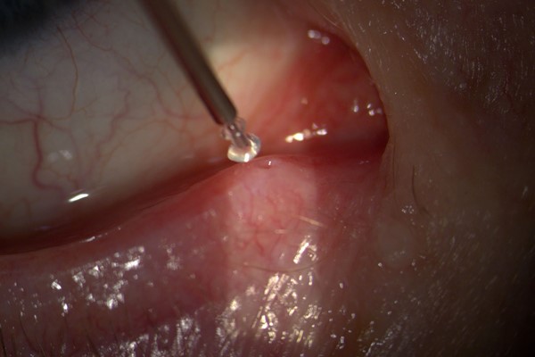
Anatomy and Mechanism
Tears normally drain from the ocular surface through the upper and lower puncta, small openings at the inner aspect of each eyelid, into the canaliculi and eventually the nasolacrimal duct and nasal cavity.
A punctal plug occludes this drainage pathway at the level of the punctum or just within the canaliculus, reducing the rate at which tears leave the ocular surface and thereby increasing the effective tear volume and residence time available to lubricate and protect the eye.
This purely mechanical mechanism is distinct from treatments that address tear production or ocular surface inflammation directly, which is why punctal plugs are often used as an adjunct alongside, rather than a replacement for, other dry eye therapies.
Types of Plugs
- Temporary, dissolvable plugs made of collagen or a similar absorbable material, lasting days to weeks, useful as a trial to predict response before committing to a longer-lasting option
- Semi-permanent silicone plugs, designed to remain in place for months to years, used once a temporary trial has confirmed meaningful symptomatic benefit
- Intracanalicular plugs, placed further within the canaliculus rather than sitting visibly at the punctal opening, offering a less visible option for patients concerned about a plug’s cosmetic appearance
All-fit Slit-Lamp Adapter
Record and share exactly what you see at the slit lamp. One adapter fits any slit lamp or surgical microscope — and any smartphone.
From Choroida — the team behind this siteIndications
Moderate to severe aqueous-deficient dry eye disease, particularly when artificial tears alone provide inadequate relief, is the classic indication for punctal plug placement.
Punctal plugs are also used to enhance the residence time of other topical medications, since reduced drainage keeps a therapeutic eye drop on the ocular surface longer, potentially improving its effect for certain chronic topical treatments.
They are generally considered after addressing any significant underlying inflammatory component of dry eye disease, since occluding drainage in an eye with substantial untreated surface inflammation can sometimes retain inflammatory mediators on the surface along with the beneficial tears.
Sequencing treatment this way, controlling inflammation first and adding punctal occlusion once that inflammatory component is reasonably settled, tends to produce a more satisfying and durable symptomatic result than occluding the puncta as a very first step.
The Trial-Before-Permanent Approach
Starting with temporary, dissolvable plugs allows both the patient and clinician to assess symptomatic response before committing to a semi-permanent silicone plug, which is a more deliberate, harder-to-reverse intervention.
This stepwise approach helps avoid placing a longer-lasting plug in a patient who either does not benefit meaningfully from punctal occlusion or who develops an unexpected side effect such as excessive tearing from the temporary trial.
Excessive tearing, or epiphora, after plug placement usually indicates either overcorrection or an underlying condition, such as significant ocular surface inflammation, that punctal occlusion alone is not addressing adequately.
Complications
Plug extrusion or migration is relatively common, particularly with punctal, rather than intracanalicular, plugs, and does not typically cause significant harm beyond simply losing the intended therapeutic effect.
Canaliculitis, discussed in relation to lacrimal disease in its own dedicated article on this site, is a recognized though uncommon complication, particularly associated with intracanalicular plugs that have migrated or become a nidus for bacterial or fungal colonization over time.
Epiphora from overcorrection is generally managed by simple plug removal, and most complications associated with punctal plugs are reversible once the plug itself is identified and addressed.
This overall reversibility is one of the more reassuring features of the treatment, and it is worth emphasizing to patients who are hesitant about trying an intervention that involves placing something into the tear drainage system.


Document what you see
Two smartphone imaging tools built for everyday clinic use — one for the slit lamp, one for the fundus.
From Choroida — the team behind this siteReferences
- Ervin AM, Law A, Pucker AD. Punctal occlusion for dry eye syndrome. Cochrane Database of Systematic Reviews.
- American Academy of Ophthalmology. Basic and Clinical Science Course, Section 8: External Disease and Cornea.
- Craig JP, Nichols KK, Akpek EK, et al. TFOS DEWS II definition and classification report. The Ocular Surface.
