
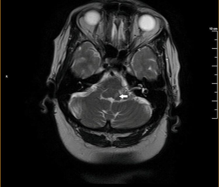
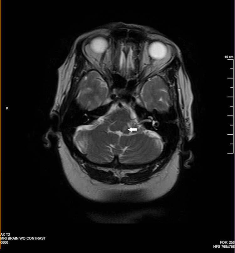
Millard-Gubler syndrome is a brainstem stroke syndrome caused by a lesion in the ventral, or basal, pons, producing ipsilateral sixth and seventh cranial nerve palsy together with contralateral body weakness, without the gaze palsy that characterizes the related Foville syndrome discussed in its own dedicated article on this site.
The two syndromes are frequently confused because they share the same ipsilateral sixth and seventh nerve findings and the same crossed pattern of contralateral weakness, but they localize to different parts of the pons, dorsal versus ventral, and that distinction is made on exam by the presence or absence of a true gaze palsy.
Getting this distinction right matters because it reflects genuinely different anatomic involvement, even though the practical clinical response, urgent stroke evaluation, is the same either way.
Like Foville syndrome, this is a genuinely uncommon presentation in everyday practice, but recognizing the pattern quickly at the bedside can meaningfully shorten the path to an accurate stroke localization.
Anatomy and Localization
The ventral pons carries the corticospinal tract on its way down to the spinal cord, along with the exiting fibers of the sixth and seventh cranial nerves as they pass through this region before emerging at the pontomedullary junction.
A lesion confined to the ventral pons, most often from occlusion of a paramedian branch of the basilar artery, damages the corticospinal tract and the exiting sixth and seventh nerve fibers together, but spares the more dorsally located paramedian pontine reticular formation responsible for horizontal gaze.
This is the precise anatomic reason Millard-Gubler syndrome lacks the gaze palsy seen in Foville syndrome: the lesion simply does not extend far enough dorsally to reach the gaze center, even though it damages structures immediately adjacent to it.
Some case descriptions also include contralateral facial sensory loss when the lesion extends slightly further to involve trigeminal fibers, though this is a variable rather than a defining feature of the syndrome.
Clinical Findings
- Ipsilateral sixth nerve palsy, causing an inability to abduct the eye on the side of the lesion
- Ipsilateral facial weakness from seventh nerve involvement, affecting both upper and lower face given the lesion’s location within the nerve’s intrapontine course
- Contralateral hemiparesis from corticospinal tract involvement, the crossed finding that, together with the ipsilateral cranial nerve findings, localizes the lesion to the brainstem
- Preserved horizontal conjugate gaze, since the pontine gaze center is not involved, distinguishing this from Foville syndrome on careful exam
All-fit Slit-Lamp Adapter
Record and share exactly what you see at the slit lamp. One adapter fits any slit lamp or surgical microscope — and any smartphone.
From Choroida — the team behind this siteDistinguishing It From Foville Syndrome
Both syndromes share ipsilateral sixth and seventh nerve palsy with contralateral hemiparesis, which is exactly why they are easy to confuse without specifically testing conjugate horizontal gaze in addition to isolated abduction of each eye.
In Millard-Gubler syndrome, the sixth nerve palsy limits abduction of the affected eye alone, while conjugate gaze toward that side, driven by the unaffected pontine gaze center, remains intact when tested with both eyes moving together.
This single test, conjugate horizontal gaze versus isolated abduction, is what separates a ventral pontine lesion from a dorsal one, and it is worth performing deliberately rather than assuming the diagnosis from the cranial nerve findings alone.
Causes
Ischemic stroke from paramedian branch occlusion of the basilar artery is the classic cause, and the same vascular risk factor profile that predicts stroke elsewhere applies here as well.
Less commonly, a pontine tumor, demyelinating lesion, or hemorrhage can produce a similar clinical picture, and the tempo of onset, sudden versus gradual, is again a useful clue for prioritizing the differential.
Evaluation and Management
Urgent MRI, preferred over CT for its superior sensitivity to brainstem pathology, is warranted in any patient with this pattern of findings, following standard acute stroke protocols when the presentation is consistent with an ischemic event.
Management is directed at the underlying cause, following the same general principles used for Foville syndrome and other brainstem stroke syndromes: acute stroke care and secondary prevention for an ischemic cause, or the appropriate pathway for a tumor or demyelinating process when imaging points elsewhere.
Multidisciplinary rehabilitation, addressing both the facial weakness and the limb weakness together, is typically needed for meaningful functional recovery after a pontine stroke of this type.
For an ophthalmologist, the practical value of knowing this syndrome is the same as for Foville syndrome: a patient presenting with an isolated-looking sixth nerve palsy who, on closer exam, also has ipsilateral facial weakness and contralateral limb weakness needs an urgent stroke workup, not a routine outpatient neuro-ophthalmology referral.


Document what you see
Two smartphone imaging tools built for everyday clinic use — one for the slit lamp, one for the fundus.
From Choroida — the team behind this siteReferences
- American Academy of Ophthalmology. Basic and Clinical Science Course, Section 5: Neuro-Ophthalmology.
- Liu GT, Volpe NJ, Galetta SL. Neuro-Ophthalmology: Diagnosis and Management.
- Kumral E, Bayulkem G, Evyapan D. Clinical spectrum of pontine infarction: clinical-MRI correlations. Journal of Neurology.
