
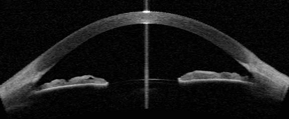
Anterior segment OCT extends optical coherence tomography, most familiar as a posterior-segment tool, to non-contact cross-sectional imaging of the cornea, angle, and anterior chamber.
It fills a genuinely different clinical niche than the posterior OCT scans most ophthalmologists use daily, and it competes with older technologies like ultrasound biomicroscopy for many of the same anterior segment questions.
Understanding where each tool actually adds value keeps a busy clinic from ordering redundant or unhelpful imaging.
Newer swept-source OCT platforms have improved anterior segment imaging depth and speed considerably compared to earlier time-domain systems, expanding what the technology can reliably capture in a single scan.
How It Differs From Posterior OCT and UBM
Anterior segment OCT uses the same basic interferometric principle as posterior OCT, but with imaging parameters optimized for the shallower depth and different reflective properties of anterior structures rather than the retina.
Unlike ultrasound biomicroscopy, which requires a water bath or gel coupling and direct probe contact with the eye, anterior segment OCT is entirely non-contact, making it faster and more comfortable, particularly for anxious or pediatric patients.
Ultrasound biomicroscopy still outperforms anterior segment OCT in imaging structures behind the iris, such as the ciliary body, since light-based OCT cannot penetrate the iris pigment epithelium the way sound waves can.
The two technologies are best thought of as complementary rather than interchangeable, with the choice between them driven by which specific structure needs to be seen rather than one uniformly replacing the other.
Clinical Uses
- Angle assessment, providing a cross-sectional, quantifiable view of angle width and configuration as a complement to gonioscopy, particularly useful for screening and for patients who cannot tolerate a goniolens comfortably
- Corneal pachymetry mapping across the entire cornea, more detailed than a single central ultrasound pachymetry reading
- Pre-refractive surgery planning, characterizing corneal thickness and shape to help exclude eyes at higher risk for post-surgical ectasia
- Monitoring corneal graft status after keratoplasty, including graft apposition, interface fluid, and early signs of rejection or detachment
- Imaging through hazy or edematous corneas where the view for a standard slit-lamp exam is degraded enough to limit direct assessment
All-fit Slit-Lamp Adapter
Record and share exactly what you see at the slit lamp. One adapter fits any slit lamp or surgical microscope — and any smartphone.
From Choroida — the team behind this siteAngle Assessment in Detail
Anterior segment OCT provides an objective, reproducible cross-sectional image of the angle that can be captured quickly and interpreted without the specific hands-on skill gonioscopy requires.
It is particularly useful as a screening tool for angle closure risk in a population setting, or for documenting angle configuration serially over time in a patient being monitored for narrowing.
Gonioscopy remains necessary for functional assessment, since it allows dynamic indentation and direct visualization of angle structures such as pigmentation and peripheral anterior synechiae that anterior segment OCT alone does not reliably capture.
The two are best used together rather than as substitutes for one another: anterior segment OCT for fast, reproducible, quantitative screening, and gonioscopy for the functional and pigmentary detail that only direct visualization provides.
Imaging an Opaque Cornea
When a cornea is too hazy or edematous for a clear slit-lamp view, anterior segment OCT can still generate a usable cross-sectional image in many cases, since it relies on a different physical principle than direct visual inspection.
This makes it valuable for assessing corneal thickness, graft-host apposition, or Descemet membrane status in an eye where the surgeon otherwise would be working somewhat blind until the cornea clears.
Intraoperative anterior segment OCT, integrated into some operating microscopes, extends this same benefit into surgery itself, letting a surgeon confirm structures such as graft orientation or residual Descemet membrane fragments in real time rather than relying on visual inspection alone.
Limitations
Image quality still depends on adequate corneal clarity, and very dense opacities or significant surface irregularity can degrade the scan enough to limit its usefulness.
Structures posterior to the iris are poorly visualized because light cannot penetrate the iris pigment epithelium, which is the specific gap that keeps ultrasound biomicroscopy clinically relevant despite anterior segment OCT’s many advantages elsewhere.
Interpretation also requires familiarity with the specific artifacts and normal variants of anterior segment scans, which differ enough from posterior segment OCT that experience with one does not automatically transfer to the other.
Cost and access remain practical constraints in many settings, since dedicated or add-on anterior segment OCT capability is not universally available the way basic slit-lamp examination and gonioscopy are.


Document what you see
Two smartphone imaging tools built for everyday clinic use — one for the slit lamp, one for the fundus.
From Choroida — the team behind this siteReferences
- Radhakrishnan S, Rollins AM, Roth JE, et al. Real-time optical coherence tomography of the anterior segment at 1310 nm. Archives of Ophthalmology.
- American Academy of Ophthalmology. Basic and Clinical Science Course, Section 8: External Disease and Cornea.
- Nolan WP, See JL, Chew PT, et al. Detection of primary angle closure using anterior segment optical coherence tomography in Asian eyes. Ophthalmology.
