
CASE REPORT
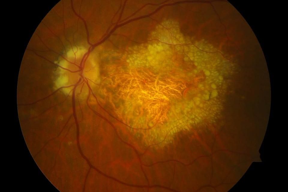
A 57‐year‐old woman presented with vision loss and distorted vision, primarily in the left eye, that had begun at the age of 47. Corrected visual acuity was 0.8 in the right eye and 0.5 in the left eye. On the left eye, metamorphopsia was reported using an Amsler grid.
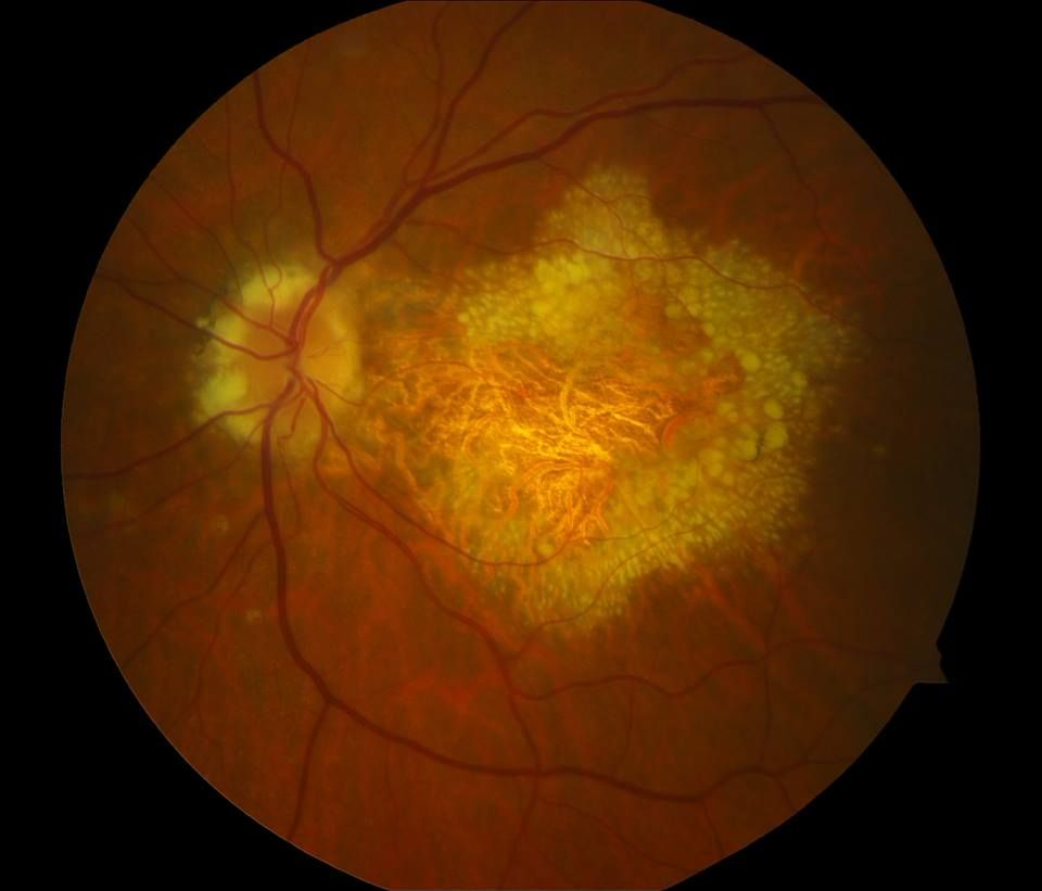
Ophthalmological examination revealed radially oriented massive hard drusen partly coalescent on both eyes in the macular region and nasal to the disc as well as macular hyperpigmentation.
Optical coherence tomography (OCT) on both eyes shows foveal drusen and retinal atrophy on the left eye. Fluorescein angiography (FLU‐A) and indocyanine green angiography (ICG‐A) showed no signs of secondary choroidal neovascularization (CNV).
The patient was clinically diagnosed with Doyne Honeycomb Retinal Dystrophy.
Doyne Honeycomb Retinal Dystrophy entity
Doyne Honeycomb Retinal Dystrophy, also known as Malattia Leventinese or Familial Dominant Drusen, all refer to the same genetic inherited retinal dystrophy characterized by an autosomal dominant mutation in the EFEMP1 gene in which patients develop early onset macular and peripapillary drusen that are often oriented radially.
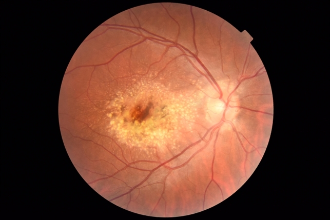
Doyne Honeycomb Retinal Dystrophy (DHRD) was first described phenotypically by Doyne in 1899 in four sisters in England. He found that each had an early onset retinal dystrophy with closely grouped white lesions in the macula and disc area which he termed the “Honeycomb” pattern.
Fundus Explorer Pro
Photograph the retinal findings described here with the phone already in your pocket — 22 D optics and built-in illumination in one handheld unit.
From Choroida — the team behind this siteDoyne Honeycomb Retinal Dystrophy management
Currently, there are no genetic or targeted therapies to correct the underlying EFEMP1 genetic mutation in DHRD. Typically, patients with DHRD are managed conservatively with observation, unless a choroidal neovascular membrane (CNVM) develops.
CNVM in DHRD is typically treated with intravitreal anti-VEGF injections. Anti-VEGF injections such as Bevacizumab have been shown to improve vision and resolve subretinal fluid.
Another treatment option is the use of lasers to clear drusen deposits. One study showed that low-energy argon laser treatment improved visual acuity and retinal sensitivity, and decreased drusen volume.
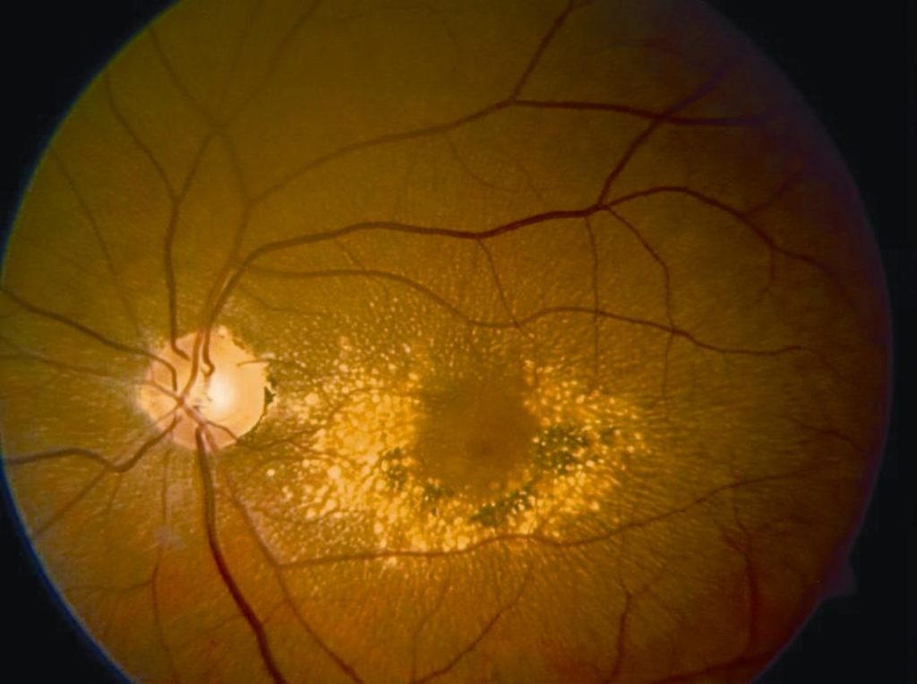
Additionally, another case report showed functional improvement using a sub-threshold retinal laser in a patient with DHRD.
Patients with a confirmed EFEMP1 mutation should have their children screened for involvement. Annual follow-up with OCT imaging in patients without CNVM is recommended.


Document what you see
Two smartphone imaging tools built for everyday clinic use — one for the slit lamp, one for the fundus.
From Choroida — the team behind this siteREFERENCES
- Doyne, R.W. Peculiar condition of choroiditis occurring in several members of the same family. Trans. Ophthalmol. Soc. UK 19, 71 –71 (1899).
- Vogt, A. in Handbuch der gesammten Augenheilkunde. Untersuchungsmethoden (eds Graefe, A. & Saemisch, T.) 1–118 (Verlag von Wilhelm Engelman, Berlin, 1925).
- Héon, Elise, et al. “Linkage of autosomal dominant radial drusen (malattia leventinese) to chromosome 2p16-21.” Archives of ophthalmology 114.2 (1996): 193-198.
- Gregory, Cheryl Y., et al. “The gene responsible for autosomal dominant Doyne’s honeycomb retinal dystrophy (DHRD) maps to chromosome 2p16.” Human molecular genetics 5.7 (1996): 1055-1059.
- Stone, Edwin M., et al. “A single EFEMP1 mutation associated with both Malattia Leventinese and Doyne honeycomb retinal dystrophy.” Nature genetics 22.2 (1999): 199-202.
- Fu, Li, et al. “The R345W mutation in EFEMP1 is pathogenic and causes AMD-like deposits in mice.” Human molecular genetics 16.20 (2007): 2411-2422.
Test yourself
A few questions straight from this article.
-
What inheritance pattern and gene underlie Doyne honeycomb retinal dystrophy?
Doyne honeycomb retinal dystrophy is an inherited retinal dystrophy caused by an autosomal dominant mutation in the EFEMP1 gene. -
Which name refers to the same condition as Doyne honeycomb retinal dystrophy?
Doyne honeycomb retinal dystrophy, Malattia Leventinese and familial dominant drusen all refer to the same EFEMP1-related retinal dystrophy. -
What drusen pattern characterizes Doyne honeycomb retinal dystrophy?
Patients develop early-onset macular and peripapillary drusen that are often radially oriented; the case showed partly coalescent radial drusen in the macula and nasal to the disc. -
In the article's Doyne honeycomb retinal dystrophy case, what did fluorescein and ICG angiography show?
Fluorescein and indocyanine green angiography showed no signs of secondary CNV, while OCT showed foveal drusen and retinal atrophy in the left eye. -
How are patients with Doyne honeycomb retinal dystrophy typically managed?
No genetic or targeted therapy corrects the EFEMP1 mutation, so patients are managed conservatively with observation unless a choroidal neovascular membrane develops. -
How is a choroidal neovascular membrane in Doyne honeycomb retinal dystrophy typically treated?
CNVM in this dystrophy is typically treated with intravitreal anti-VEGF injections; bevacizumab has been shown to improve vision and resolve subretinal fluid. -
What did one study report after low-energy argon laser in Doyne honeycomb retinal dystrophy?
Low-energy argon laser improved visual acuity and retinal sensitivity and decreased drusen volume; a separate case report found functional improvement with sub-threshold laser. -
What follow-up is recommended for Doyne honeycomb retinal dystrophy patients without a neovascular membrane?
Annual follow-up with OCT imaging is recommended for patients who have not developed a choroidal neovascular membrane. -
What does the article advise for the children of a Doyne honeycomb dystrophy patient with a confirmed EFEMP1 mutation?
Patients with a confirmed EFEMP1 mutation should have their children screened for involvement, as the condition is inherited as an autosomal dominant trait.
