
Dominant optic atrophy (Kjer type) is the most common inherited optic neuropathy overall, caused in the large majority of cases by mutations in OPA1, a gene encoding a mitochondrial protein essential for normal mitochondrial fusion and function within retinal ganglion cells.
Unlike Leber hereditary optic neuropathy, its mitochondrially inherited counterpart discussed in its own dedicated article on this site, dominant optic atrophy follows autosomal dominant inheritance, typically presents earlier and more insidiously, and generally produces a milder, though still lifelong and functionally significant, degree of visual impairment.
Why Mitochondrial Function Matters to the Optic Nerve
Retinal ganglion cells, and their unmyelinated axons within the retina before they reach the optic nerve, are unusually dependent on efficient mitochondrial energy production.
This is why a disruption of mitochondrial dynamics — even one present throughout the entire body, as OPA1 mutations are — manifests clinically with such striking selectivity for this one cell population.
This shared vulnerability of retinal ganglion cell mitochondria is also the conceptual link between dominant optic atrophy and Leber hereditary optic neuropathy, despite their different inheritance patterns and genetic causes.
Clinical Presentation
The disease typically presents insidiously in early childhood, often before age ten, with slowly progressive, bilateral (though sometimes asymmetric) vision loss that is frequently mild enough in its early stages to go unnoticed by the child or family.
Many cases are only picked up on a routine school vision screening or incidental eye exam rather than because of a specific visual complaint.
Because the onset is so gradual and the disease congenital in its underlying genetic basis, some patients and families are unaware that their vision is reduced relative to normal, having had no other baseline for comparison.
Fundus Explorer Pro
Photograph the retinal findings described here with the phone already in your pocket — 22 D optics and built-in illumination in one handheld unit.
From Choroida — the team behind this siteClinical Findings
- Bilateral, gradually progressive, generally mild to moderate reduction in visual acuity, typically in a range that preserves substantial functional vision for most patients rather than causing severe or profound loss
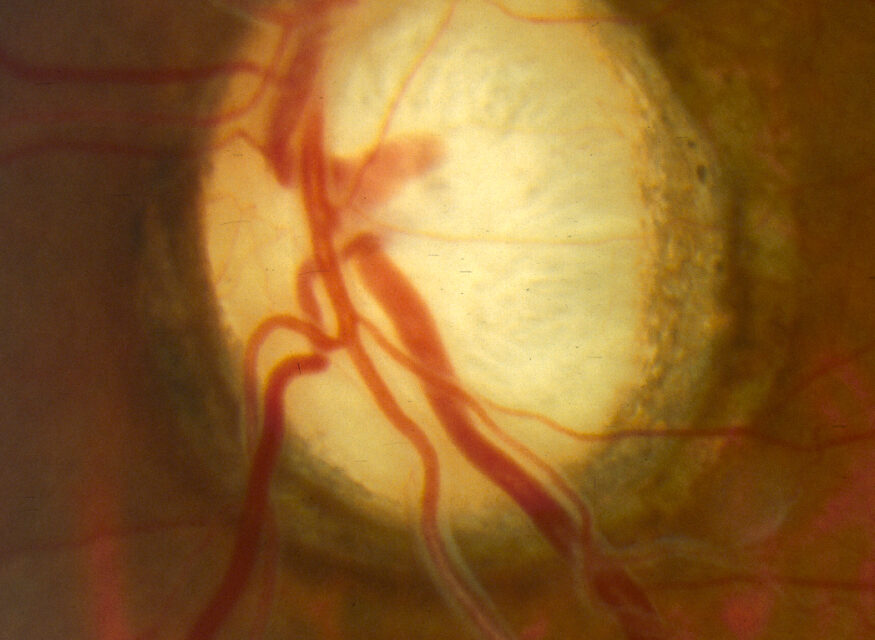
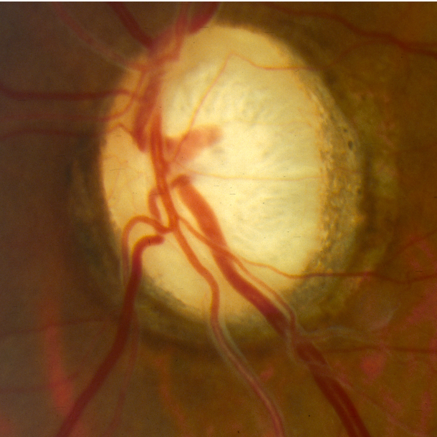
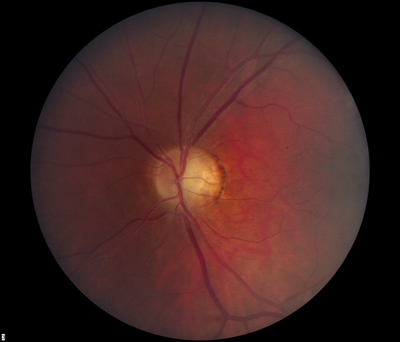
- Optic disc pallor, classically most pronounced in the temporal quadrant, corresponding to preferential loss of the papillomacular bundle
- Color vision deficits, particularly a blue-yellow (tritanopic) pattern, which is somewhat distinctive since most other optic neuropathies more typically produce red-green deficits — this tritanopic pattern is a useful, if not entirely specific, clinical clue supporting the diagnosis
- Central, cecocentral, or paracentral visual field defects on formal testing
- Significant variability in severity even within the same family, since OPA1-related disease shows notably variable penetrance and expressivity, meaning some family members can be quite mildly affected while others in the same pedigree have more significant vision loss
Syndromic (OPA1-Plus) Disease
A subset of patients with specific OPA1 mutations develop additional extraocular features beyond isolated optic atrophy — sensorineural hearing loss, peripheral neuropathy, chronic progressive external ophthalmoplegia, and myopathy — a pattern sometimes referred to as “DOA plus” or syndromic dominant optic atrophy.
Recognizing this broader syndromic possibility is worthwhile once the diagnosis of dominant optic atrophy is established, because it can prompt additional relevant screening (audiology, in particular) that isolated optic atrophy alone would not.
Differential Diagnosis
- Leber hereditary optic neuropathy — mitochondrial inheritance, typically more acute or subacute onset with sequential eye involvement, and generally more severe vision loss than dominant optic atrophy, discussed in its own dedicated article on this site
- Toxic and nutritional optic neuropathy — an acquired rather than inherited cause of bilateral, symmetric optic neuropathy, distinguished by a relevant exposure or dietary history and typically a later age of onset, covered in its own dedicated article
- Bilateral compressive optic neuropathy — from a chiasmal or bilateral optic nerve mass lesion, generally showing a distinct field defect pattern (such as bitemporal hemianopia with chiasmal lesions) and warranting neuroimaging to exclude
- Other inherited optic neuropathies and mitochondrial syndromes with overlapping features, distinguished through targeted genetic testing when the clinical picture and family history do not clearly fit the classic dominant optic atrophy pattern
Diagnostic Evaluation
A three-generation family history, looking specifically for other relatives with unexplained mild vision loss (who may never have been formally diagnosed), is a valuable and often underused diagnostic tool given the variable expressivity of the condition.
Genetic testing for OPA1 mutations confirms the diagnosis and is increasingly accessible, useful both for confirming the clinical impression and for family counseling regarding the autosomal dominant inheritance pattern and its associated 50 percent transmission risk to offspring of an affected individual.
Management
There is currently no treatment that reverses or halts the underlying mitochondrial dysfunction driving dominant optic atrophy.
Management centers on low vision support, appropriate educational accommodations for affected children, and genetic counseling for the family, with the specific accommodations tailored to the individual child’s actual level of visual function rather than a generic plan.
Ongoing monitoring is reasonable given the variable natural history, since documenting a stable baseline allows any unexpected acceleration in vision loss to be recognized promptly rather than attributed automatically to the expected course of the disease.
Patients with syndromic features should be referred for the relevant additional specialty evaluation (audiology for hearing loss, neurology for peripheral neuropathy or myopathy) as appropriate to their specific presentation, ensuring that a purely ophthalmic diagnosis does not become a barrier to recognizing broader systemic involvement.


Document what you see
Two smartphone imaging tools built for everyday clinic use — one for the slit lamp, one for the fundus.
From Choroida — the team behind this siteReferences
- Yu-Wai-Man P, Griffiths PG, Chinnery PF. Mitochondrial optic neuropathies — disease mechanisms and therapeutic strategies. Progress in Retinal and Eye Research.
- Cohn AC, Toomes C, Potter C, et al. Autosomal dominant optic atrophy: penetrance and expressivity in patients with OPA1 mutations. American Journal of Ophthalmology.
- American Academy of Ophthalmology. Basic and Clinical Science Course, Section 5: Neuro-Ophthalmology.
