
A nutritional deficiency that costs almost nothing to prevent remains one of the world’s leading causes of preventable childhood blindness.
Keratomalacia is the end-stage corneal complication of severe vitamin A deficiency, and it can destroy a cornea within days once it begins.
The disease follows a graded progression — xerophthalmia — from simple night blindness through conjunctival and corneal drying to frank corneal melting.
Each stage is a distinct, recognizable clinical entity, and catching the disease early is the difference between full recovery and permanent blindness.
It remains a significant problem in regions with limited dietary vitamin A intake, disproportionately affecting young children.
Recognizing the early ocular surface signs — well before the cornea itself is threatened — is what actually prevents this form of blindness.
What Is Keratomalacia?
Keratomalacia is corneal liquefactive necrosis occurring as the most severe stage of xerophthalmia, the spectrum of ocular manifestations of vitamin A deficiency.
The WHO xerophthalmia classification stages the disease:
- XN — night blindness, the earliest functional sign
- X1A — conjunctival xerosis (dryness)
- X1B — Bitot spots, foamy keratinized patches on the bulbar conjunctiva
- X2 — corneal xerosis
- X3A/X3B — corneal ulceration/keratomalacia involving less than or at least one-third of the corneal surface
- XS — corneal scar; XF — xerophthalmic fundus changes
Keratomalacia (X3) represents the point at which the disease becomes acutely sight-threatening, rather than simply visible on examination.
Epidemiology
Vitamin A deficiency remains a significant global public health problem, concentrated in specific vulnerable populations.
- Most severe cases occur in young children in regions with limited dietary vitamin A intake
- It is a leading preventable cause of childhood blindness in low-resource settings
- Measles infection is a well-recognized precipitant of acute vitamin A deficiency and keratomalacia in at-risk children
- Malnutrition, chronic diarrheal illness, and poor weaning practices all compound risk in endemic regions
Because keratomalacia can progress from mild ocular surface drying to corneal destruction within days in a severely deficient child, it represents a genuine pediatric emergency wherever it is suspected.
All-fit Slit-Lamp Adapter
Record and share exactly what you see at the slit lamp. One adapter fits any slit lamp or surgical microscope — and any smartphone.
From Choroida — the team behind this sitePathophysiology
Vitamin A is essential for maintaining normal epithelial differentiation throughout the ocular surface.
- Vitamin A deficiency impairs normal mucin-producing goblet cell function and conjunctival epithelial differentiation
- The conjunctiva undergoes squamous metaplasia and keratinization, producing the foamy appearance of Bitot spots
- Reduced tear film mucin and instability lead to progressive corneal surface drying (xerosis)
- Severe, prolonged deficiency triggers corneal epithelial breakdown and stromal liquefactive necrosis — keratomalacia
- Concurrent systemic illness (particularly measles) and protein-energy malnutrition accelerate this progression dramatically
Because the underlying process is a systemic nutritional deficiency, the eye findings are really a visible marker of a body-wide problem, not an isolated ocular disease.
Risk Factors
Nutritional Risk Factors
- Diets low in vitamin A or its precursors (limited animal-source foods, dairy, and orange/dark green vegetables)
- Protein-energy malnutrition, which compounds vitamin A deficiency
- Early cessation of breastfeeding without adequate complementary vitamin A intake
Precipitating and Amplifying Factors
- Measles infection, a classic and well-documented trigger for acute deterioration
- Chronic diarrheal disease, which impairs vitamin A absorption
- Other conditions causing fat malabsorption, since vitamin A is fat-soluble
A child presenting with keratomalacia in the setting of recent measles is a recognized, high-risk clinical pattern that should prompt urgent systemic as well as ocular treatment.
Clinical Presentation
Symptoms
- Difficulty seeing in dim light or at night (nyctalopia), often the earliest reported symptom
- Ocular dryness, irritation, and foreign-body sensation as conjunctival and corneal xerosis develop
- Rapid onset of pain, photophobia, and visible corneal clouding once keratomalacia begins
- Systemic signs of malnutrition or recent measles infection in many affected children
Examination Findings
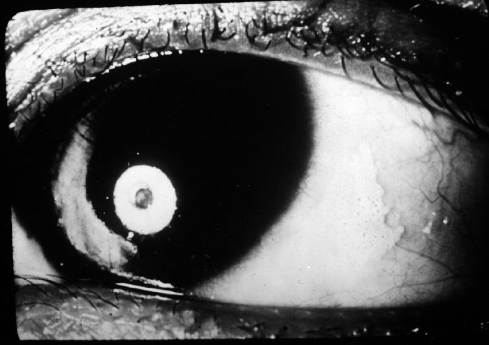
- Bitot spots — foamy, triangular, keratinized patches typically on the temporal bulbar conjunctiva
- A dry, non-wettable, roughened conjunctival and corneal surface (xerosis)
- Corneal haze, ulceration, or frank melting and perforation in advanced keratomalacia
- Bilateral involvement is typical, reflecting the systemic nature of the deficiency
Bitot spots are a highly specific, easily recognized bedside sign — finding them in a malnourished child should immediately prompt vitamin A treatment, not just observation.
Diagnostic Evaluation
Clinical Staging
- Diagnosis and staging are primarily clinical, using the WHO xerophthalmia classification
- A history of dietary intake, recent measles, and diarrheal illness supports the diagnosis
Laboratory and Systemic Assessment
- Serum retinol levels can confirm deficiency where available, though treatment in a clinically consistent presentation should not wait for lab confirmation
- Assessment for coexisting protein-energy malnutrition and systemic illness, since these commonly occur together
In a child with the classic clinical picture, treatment should begin immediately — keratomalacia can progress from mild xerosis to corneal perforation over a matter of days.
Differential Diagnosis
Conditions that can resemble xerophthalmia include:
- Severe dry eye disease from other causes — lacks the systemic nutritional context and Bitot spots
- Infectious keratitis (bacterial, fungal, herpetic) — typically unilateral and without conjunctival xerosis or Bitot spots
- Trachoma — conjunctival scarring and trichiasis rather than the foamy xerosis of vitamin A deficiency
- Stevens-Johnson syndrome/toxic epidermal necrolysis — severe ocular surface disease with a clear preceding drug or infectious trigger and mucocutaneous involvement
The combination of Bitot spots, bilateral conjunctival xerosis, and a malnourished or recently measles-infected child is distinctive enough to separate xerophthalmia from these mimics.
Management
Emergency Vitamin A Supplementation
- High-dose oral vitamin A given immediately on diagnosis, with a repeat dose the following day, and a third dose two to four weeks later, per WHO protocol
- Treatment should begin as soon as the diagnosis is suspected clinically, without waiting for laboratory confirmation
Ocular Surface Care
- Topical lubrication and antibiotic prophylaxis to protect a compromised ocular surface from secondary infection
- Careful monitoring for corneal perforation, which may require urgent surgical intervention (tissue adhesive, amniotic membrane, or emergency keratoplasty) in advanced cases
Systemic and Public Health Measures
- Treatment of any concurrent measles infection, malnutrition, or diarrheal illness
- Community-level vitamin A supplementation programs and dietary diversification are the primary tools for preventing new cases
- Measles vaccination programs indirectly reduce keratomalacia risk by preventing a major precipitating illness
Public health prevention — supplementation programs and measles vaccination — has done far more to reduce global keratomalacia than any treatment given after the disease has already started.
Prognosis
Outcomes depend almost entirely on how early treatment begins.
- Night blindness and early conjunctival xerosis (XN, X1A, X1B) resolve completely with prompt vitamin A treatment
- Corneal xerosis and early ulceration (X2, X3A) can also recover well if treated before extensive tissue loss
- Established keratomalacia with significant corneal destruction (X3B) frequently leaves permanent scarring or results in blindness despite treatment
Because the window between reversible xerosis and irreversible corneal destruction can be just days, early recognition — not advanced treatment technology — is what actually saves vision in this disease.


Document what you see
Two smartphone imaging tools built for everyday clinic use — one for the slit lamp, one for the fundus.
From Choroida — the team behind this siteReferences
- World Health Organization. Global prevalence of vitamin A deficiency in populations at risk 1995-2005: WHO global database on vitamin A deficiency.
- Sommer A. Vitamin A deficiency and clinical disease: an historical overview. Journal of Nutrition. 2008.
- WHO/UNICEF. Indicators for assessing vitamin A deficiency and their application in monitoring and evaluating intervention programmes.
- Keratomalacia. StatPearls, NCBI Bookshelf.
- Xerophthalmia. EyeWiki, American Academy of Ophthalmology.
Test yourself
A few questions straight from this article.
-
Which nutritional deficiency causes keratomalacia and the wider spectrum of xerophthalmia?
Keratomalacia is the end-stage corneal complication of severe vitamin A deficiency, and the ocular signs are a visible marker of a body-wide nutritional problem. -
What is the earliest functional sign of vitamin A deficiency in the xerophthalmia sequence?
Night blindness (XN) is the first stage of the WHO xerophthalmia classification and is usually the earliest symptom a patient or parent reports. -
In the WHO xerophthalmia classification, which stage corresponds to Bitot spots?
The WHO scale runs XN (night blindness), X1A (conjunctival xerosis), X1B (Bitot spots), X2 (corneal xerosis) and X3A/X3B (ulceration/keratomalacia). -
What distinguishes stage X3B from X3A keratomalacia in the WHO classification?
X3A denotes corneal ulceration or keratomalacia affecting less than one-third of the cornea; X3B involves at least one-third, and it is the stage that most often ends in blindness. -
What produces the foamy appearance of Bitot spots in vitamin A deficiency?
Vitamin A deficiency disrupts goblet cell function and epithelial differentiation, so the conjunctiva keratinizes and forms the foamy, triangular keratinized patch known as a Bitot spot. -
Where are Bitot spots most typically found in a child with vitamin A deficiency?
Bitot spots are foamy, triangular, keratinized patches classically sited on the temporal bulbar conjunctiva, and finding them should prompt immediate vitamin A treatment. -
Which systemic infection is a classic precipitant of acute keratomalacia in at-risk children?
Measles is a well-recognized trigger for acute deterioration in vitamin A deficiency, which is why measles vaccination programs indirectly reduce keratomalacia. -
How does keratomalacia usually present in terms of laterality?
Because the underlying deficiency is systemic, keratomalacia is typically bilateral, which helps separate it from unilateral infectious keratitis. -
How should vitamin A treatment be timed in a child with clinically suspected keratomalacia?
High-dose oral vitamin A is given on diagnosis, repeated the next day and again two to four weeks later; serum retinol confirmation should never delay the first dose. -
Which xerophthalmia stage most often leaves permanent corneal scarring or blindness despite treatment?
Night blindness and early xerosis resolve completely with prompt vitamin A, and X2/X3A can still recover, but established X3B destruction frequently leaves permanent scarring.
