
Case Study
A 34-year-old male was referred for a routine ophthalmic examination after noticing mild night vision difficulty without significant central visual loss.

He had no history of ocular inflammation, trauma, or systemic disease. Best-corrected visual acuity was 20/20 in both eyes.
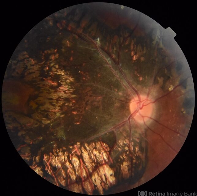
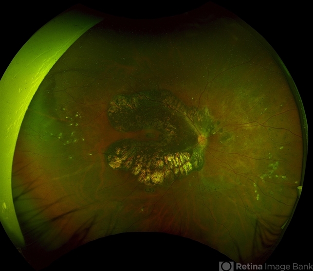
Fundus examination revealed bilateral, symmetric areas of chorioretinal atrophy with bone-spicule–like pigmentation distributed along the retinal veins, extending from the posterior pole toward the midperiphery.
The macula was relatively spared. These characteristic findings led to a diagnosis of pigmented paravenous retinochoroidal atrophy (PPRCA).
Disease Entity
Pigmented paravenous retinochoroidal atrophy (PPRCA) is a rare, typically bilateral retinal disorder characterized by zones of retinal pigment epithelium (RPE) and chorioretinal atrophy distributed along the retinal veins.
The condition is usually discovered incidentally and often follows a benign, slowly progressive course.
PPRCA is considered a distinct clinical entity, although its exact etiology remains unclear.
It is generally regarded as non-inflammatory and non-hereditary, with most cases occurring sporadically.
Fundus Explorer Pro
Photograph the retinal findings described here with the phone already in your pocket — 22 D optics and built-in illumination in one handheld unit.
From Choroida — the team behind this sitePathophysiology
The underlying mechanism of PPRCA is not fully understood. Several hypotheses have been proposed, including:
-
Developmental abnormalities of the retinal pigment epithelium
-
Previous subclinical inflammatory insult affecting paravenous regions
-
Primary choroidal or retinal vascular dysfunction
Histopathologic changes primarily involve:
-
Degeneration of the RPE
-
Secondary photoreceptor loss
-
Choriocapillaris atrophy in advanced areas
The paravenous distribution suggests a possible relationship to venous drainage patterns or localized metabolic susceptibility.
Epidemiology
-
Extremely rare condition
-
Typically diagnosed in young to middle-aged adults
-
No strong gender predilection
-
Usually bilateral and symmetric
-
Often asymptomatic at presentation
Familial cases are rare, and no consistent genetic association has been identified.
Clinical Features
Most patients with PPRCA are asymptomatic or have mild visual complaints.
Possible symptoms include:
-
Mild nyctalopia
-
Subtle peripheral visual field defects
-
Rarely decreased central vision in advanced cases
Central visual acuity is usually preserved due to macular sparing.

Examination Findings
Fundus Examination
-
Linear or branching areas of chorioretinal atrophy along retinal veins
-
Bone-spicule–like pigmentation confined to paravenous regions
-
Sharply demarcated atrophic zones
-
Relative sparing of the macula and optic disc
Imaging
-
Fundus autofluorescence: Hypoautofluorescent paravenous streaks
-
OCT: Thinning of outer retinal layers and RPE loss along affected veins
-
Fluorescein angiography: Window defects without active leakage
These imaging features help distinguish PPRCA from inflammatory or hereditary retinopathies.
Differential Diagnosis
-
Retinitis pigmentosa
-
Healed retinal vasculitis
-
Serpiginous choroiditis
-
Choroideremia
-
Perivenous atrophy secondary to inflammatory disease
The absence of active inflammation and the characteristic paravenous pattern are key differentiating features.
Diagnosis
Diagnosis is primarily clinical, supported by multimodal imaging. A thorough history is essential to exclude prior uveitis, retinal vasculitis, or infectious etiologies. Laboratory testing is not routinely required unless inflammatory disease is suspected.
Management
There is no specific treatment for PPRCA.
Management focuses on:
-
Patient reassurance
-
Periodic follow-up to monitor progression
-
Visual field testing in symptomatic patients
In most cases, the disease remains stable over time.
Prognosis
The visual prognosis of PPRCA is generally favorable.
-
Slow or minimal progression
-
Central vision typically preserved
-
Severe visual impairment is uncommon
Long-term follow-up studies suggest that many patients maintain good functional vision throughout life.


Document what you see
Two smartphone imaging tools built for everyday clinic use — one for the slit lamp, one for the fundus.
From Choroida — the team behind this siteReferences
-
Franceschetti A. Pigmented paravenous retinochoroidal atrophy. Arch Ophthalmol.
-
Shields JA, Shields CL. Intraocular Tumors: An Atlas and Textbook.
-
Yanoff M, Duker JS. Ophthalmology. 5th ed. Elsevier.
-
American Academy of Ophthalmology. Basic and Clinical Science Course: Retina and Vitreous.
-
Tasman W, Jaeger EA. Duane’s Ophthalmology.
Test yourself
A few questions straight from this article.
-
What defines pigmented paravenous retinochoroidal atrophy (PPRCA)?
PPRCA is a rare, usually bilateral disorder in which zones of RPE and chorioretinal atrophy are distributed along the retinal veins. -
How is the etiology of pigmented paravenous retinochoroidal atrophy generally regarded?
PPRCA is generally regarded as non-inflammatory and non-hereditary, with most cases sporadic; familial cases are rare and no consistent genetic association is known. -
Which histopathologic change is central to pigmented paravenous retinochoroidal atrophy?
The changes primarily involve RPE degeneration with secondary photoreceptor loss, plus choriocapillaris atrophy in advanced areas. -
Which age group is pigmented paravenous retinochoroidal atrophy typically diagnosed in?
PPRCA is extremely rare and is typically diagnosed in young to middle-aged adults, with no strong gender predilection and frequent incidental discovery. -
Which symptom may patients with pigmented paravenous retinochoroidal atrophy report?
Most patients are asymptomatic, but mild nyctalopia and subtle peripheral field defects may occur; central acuity is usually preserved because the macula is spared. -
Which fundus autofluorescence pattern is seen in pigmented paravenous retinochoroidal atrophy?
Fundus autofluorescence shows hypoautofluorescent streaks following the paravenous distribution of RPE loss. -
What does fluorescein angiography show in pigmented paravenous retinochoroidal atrophy?
FA demonstrates window defects corresponding to areas of RPE atrophy, with no active leakage — a finding that helps exclude inflammatory disease. -
Which two features best separate PPRCA from an inflammatory retinopathy?
The article names the absence of active inflammation together with the characteristic paravenous distribution as the key differentiating features from retinitis pigmentosa, healed vasculitis and similar entities. -
What is the recommended management of pigmented paravenous retinochoroidal atrophy?
There is no specific treatment; management centres on patient reassurance, periodic follow-up for progression and visual field testing in symptomatic patients. -
What is the expected visual prognosis in pigmented paravenous retinochoroidal atrophy?
The prognosis is generally favourable: progression is slow or minimal, central vision is typically preserved and severe visual impairment is uncommon.
