
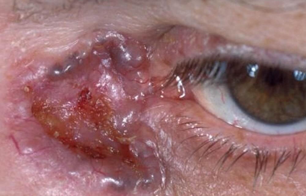
Basal cell carcinoma is the most common malignant tumor of the eyelid by a wide margin, accounting for the large majority of all eyelid malignancies.
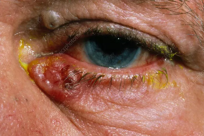
While its metastatic potential is low, it is locally aggressive and can cause significant tissue destruction — including orbital invasion — if allowed to grow unchecked, which is why timely diagnosis and treatment matter even for a tumor that is rarely, if ever, going to spread to distant sites.
This local-only pattern of aggression is a useful way to frame the stakes for patients: the danger is not death from metastasis, in the great majority of cases, but the real and cumulative risk of progressive, disfiguring, functionally significant tissue loss around the eye if the tumor is allowed to continue growing untreated.
Risk Factors
- Cumulative ultraviolet sun exposure — the dominant risk factor, consistent with the lower eyelid (which receives more direct sun exposure than the upper lid) being the single most common site of occurrence
- Fair skin, light eye color, and a tendency to sunburn rather than tan
- Older age
- Immunosuppression
- A history of prior basal cell carcinoma, which substantially raises the risk of additional lesions developing elsewhere
- Basal cell nevus (Gorlin) syndrome — a genetic condition producing multiple basal cell carcinomas at a young age, alongside other developmental abnormalities, and worth considering in a young patient with an eyelid basal cell carcinoma or with multiple lesions
Clinical Patterns
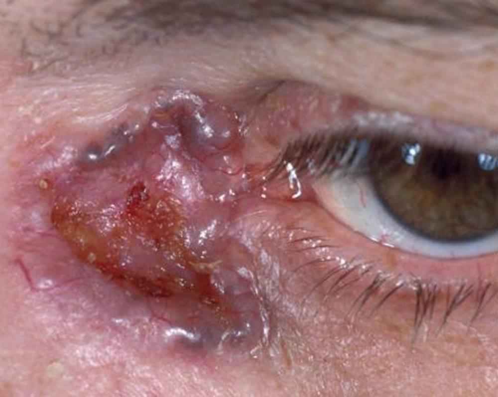
- Nodular — the most common pattern, a pearly, translucent papule or nodule with overlying telangiectatic vessels, often with a central area of ulceration (rodent ulcer) in more established lesions
- Morpheaform (sclerosing) — a flatter, scar-like, indurated plaque with poorly defined margins, notoriously more aggressive and more difficult to fully excise given how much the true extent of the tumor is underestimated by its visible borders
- Superficial — a scaly, erythematous, plaque-like lesion, less common on the eyelid than the more classic nodular pattern
The morpheaform variant deserves particular attention because its indistinct, ill-defined clinical margins so often extend well beyond what is visible.
This is a major reason Mohs micrographic surgery has become the preferred approach for eyelid basal cell carcinoma generally, and especially for this pattern.
All-fit Slit-Lamp Adapter
Record and share exactly what you see at the slit lamp. One adapter fits any slit lamp or surgical microscope — and any smartphone.
From Choroida — the team behind this siteClinical Presentation
Patients typically present with a slow-growing eyelid lesion, often present for months before seeking evaluation given how gradual and painless the growth usually is, most commonly on the lower eyelid or medial canthus.
Lash loss (madarosis) over the lesion, distortion of the normal lid margin architecture, and a lesion that bleeds easily or fails to heal are all clinical clues supporting malignancy over a benign process.
A lesion that patients describe as a “pimple that never fully heals” or that periodically scabs over and bleeds again is a particularly characteristic history, and should prompt biopsy rather than continued reassurance or empiric treatment for a presumed benign cause.
Differential Diagnosis
- Sebaceous carcinoma — can mimic chalazion or chronic blepharitis, discussed in its own dedicated article on this site, with a notably worse metastatic potential than basal cell carcinoma despite sometimes similar initial appearance
- Squamous cell carcinoma — can appear similarly, with a somewhat higher metastatic potential than basal cell carcinoma, definitively distinguished on histopathology
- Benign lesions — chalazion, papilloma, seborrheic keratosis, and intradermal nevus can all mimic early or atypical basal cell carcinoma, particularly before the classic pearly, telangiectatic appearance has fully developed
- Actinic keratosis — a premalignant lesion in its own right, occurring on the same sun-exposed skin and worth distinguishing since management differs
Diagnostic Evaluation
Biopsy confirms the diagnosis and histologic subtype, information that directly informs the surgical approach.
A well-defined nodular lesion may be managed differently than an infiltrative or morpheaform pattern given their different tendencies toward subclinical extension.
Imaging (CT or MRI) is reserved for larger or more advanced lesions with clinical concern for orbital invasion, which, though uncommon, represents the most feared local complication of untreated or inadequately treated disease, since orbital involvement significantly raises both the surgical complexity and the risk to the globe.
Management
Mohs micrographic surgery has become the preferred treatment for most eyelid basal cell carcinomas, offering the highest cure rates of any treatment modality while sparing as much healthy surrounding tissue as possible.
This is an important consideration given the limited amount of eyelid tissue available for reconstruction and the functional and cosmetic stakes of eyelid surgery specifically, where even a small amount of unnecessarily sacrificed tissue can meaningfully affect the final functional and cosmetic outcome.
Standard surgical excision with frozen-section margin control is a reasonable alternative where Mohs surgery is not available, though generally with somewhat lower reported cure rates than Mohs, particularly for higher-risk histologic subtypes.
Reconstruction of the resulting defect, tailored to its size and location, is often needed to restore both eyelid function (protecting the ocular surface) and cosmesis, and is typically planned in coordination with, or immediately following, the excisional procedure.
Radiation therapy and topical or systemic hedgehog pathway inhibitors (vismodegib, sonidegib) are reserved for patients who are not surgical candidates or for locally advanced or recurrent disease not amenable to further surgery, and their use is generally coordinated closely with dermatology or oncology given the systemic nature of these targeted agents.


Document what you see
Two smartphone imaging tools built for everyday clinic use — one for the slit lamp, one for the fundus.
From Choroida — the team behind this siteReferences
- Cook BE Jr, Bartley GB. Treatment options and future prospects for the management of eyelid malignancies. Ophthalmology.
- Margo CE, Waltz K. Basal cell carcinoma of the eyelid and periocular skin. Survey of Ophthalmology.
- American Academy of Ophthalmology. Basic and Clinical Science Course, Section 7: Orbit, Eyelids, and Lacrimal System.
