
Pellucid marginal degeneration is a rare, bilateral, non-inflammatory corneal ectasia distinguished by a band of inferior peripheral thinning that sits below and is separated from the area of maximal corneal protrusion.
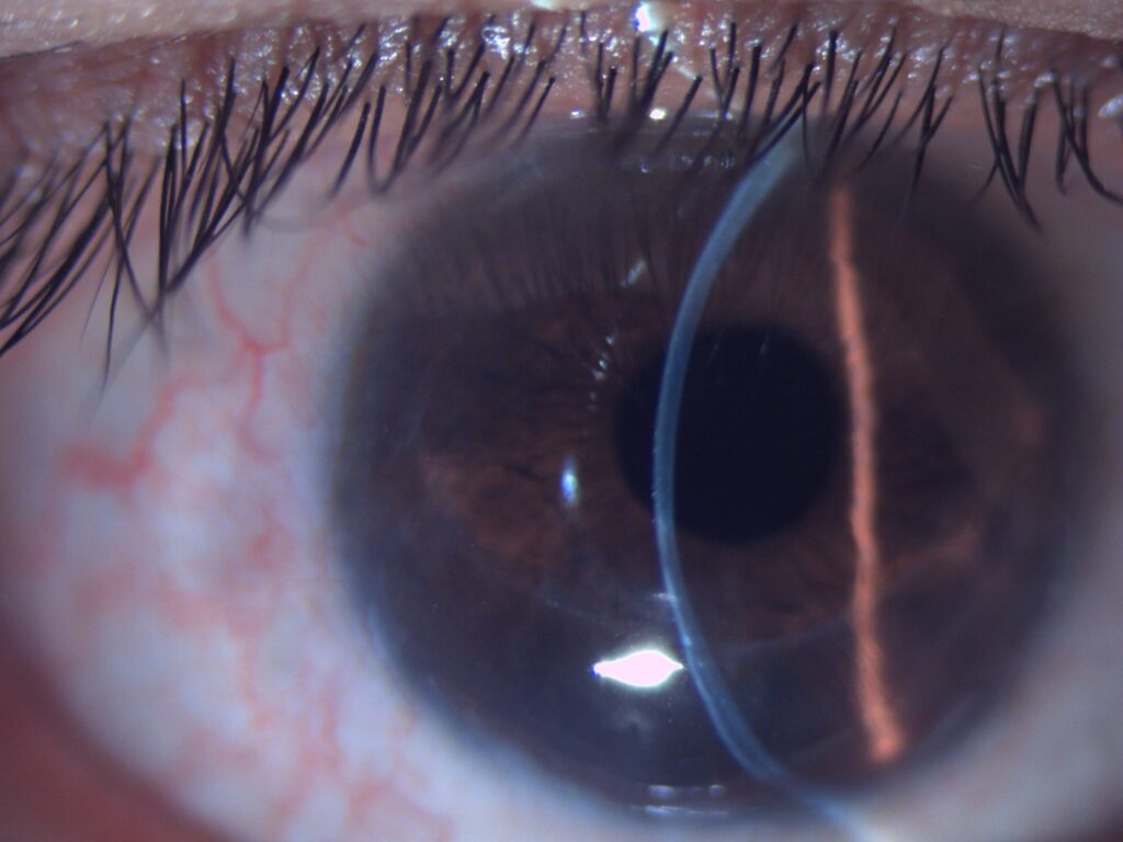
This pattern produces one of the most distinctive topographic signatures in all of corneal disease and can look confusingly similar to keratoconus without careful attention to exactly where the thinning and the steepening are each located.
Correctly recognizing the difference matters practically, since the two conditions are managed with somewhat different contact lens fitting strategies and, when surgery is needed, genuinely different surgical approaches.
What Makes It Different From Keratoconus
In keratoconus, the zone of maximal thinning and the zone of maximal protrusion (the cone apex) are the same location, typically central or paracentral.
In pellucid marginal degeneration, the thinning is confined to a narrow, crescentic band in the inferior periphery (typically between the four and eight o’clock positions, a few millimeters from the limbus), while the cornea actually protrudes and steepens above this thinned band, closer to the visual axis.
The ectasia is, in effect, displaced from the thinning itself, mechanically and topographically distinct from keratoconus even though both conditions ultimately cause irregular astigmatism from progressive corneal ectasia.
This spatial separation between the thinnest point and the steepest point is the single feature that a careful topographic review should focus on whenever the two conditions are being distinguished, since relying on the general clinical impression of “an ectatic-looking cornea” alone is not sufficient to tell them apart.
Clinical Presentation
Patients typically present in the third to fifth decade — later than the more classic teenage-to-young-adult onset of keratoconus — with gradually progressive, high, irregular astigmatism and reduced best-corrected vision from the irregular corneal surface.
Unlike keratoconus, acute hydrops is rare in pellucid marginal degeneration, because the thinning is peripheral rather than central and the mechanical dynamics of the cornea differ in a way that makes sudden Descemet’s membrane rupture much less common.
Because the condition is typically diagnosed later in life than keratoconus, patients are more likely to have already been wearing glasses or soft contact lenses for years, and progressively worsening or increasingly difficult-to-correct astigmatism in an established, previously stable prescription is often what first draws attention to the possibility of an underlying ectasia.
All-fit Slit-Lamp Adapter
Record and share exactly what you see at the slit lamp. One adapter fits any slit lamp or surgical microscope — and any smartphone.
From Choroida — the team behind this siteExam and Imaging Findings
- A thin, clear (avascular, non-inflamed, non-opaque) band of inferior peripheral corneal thinning — the term “pellucid” specifically refers to this clear, translucent quality of the thinned band
- Protrusion and steepening of the cornea above the thinned band, closer to but usually not involving the central visual axis directly
- Corneal topography showing a characteristic “crab claw” or “butterfly” pattern of inferior against-the-rule astigmatism, one of the most recognizable topographic signatures in corneal imaging
- Absence of the central or paracentral apical thinning and Vogt’s striae typical of keratoconus
- Progressive against-the-rule astigmatism over time, which is itself a distinguishing clinical clue since keratoconus more typically produces with-the-rule or oblique astigmatism
Differential Diagnosis
- Keratoconus — central or paracentral thinning and protrusion at the same location, more typically with-the-rule astigmatism, earlier onset, higher hydrops risk
- Keratoglobus — diffuse, generalized corneal thinning extending to the limbus, rather than a localized inferior band
- Terrien’s marginal degeneration — peripheral thinning, but typically superior rather than inferior, in older patients, and associated with mild inflammation and lipid deposition that pellucid marginal degeneration lacks
- Inferior keratoconus — a variant of keratoconus with inferior displacement of the cone, which can be topographically confused with pellucid marginal degeneration and requires careful distinction based on the exact relationship between thinning and steepening zones
Diagnostic Evaluation
Corneal topography is the essential diagnostic tool, since the characteristic crab-claw or butterfly pattern is far more reliably identified on a topographic map than on slit-lamp exam alone, particularly in earlier, more subtle cases.
Corneal tomography, which adds posterior corneal surface and pachymetric information to the anterior topographic map, further improves detection of subtle or early disease and helps distinguish pellucid marginal degeneration from inferior keratoconus when the pattern is ambiguous.
Serial topography over time documents whether the ectasia is progressing, information that directly affects decisions about contact lens fitting, the timing of any consideration of collagen cross-linking, and long-term monitoring frequency.
Management
Spectacle correction manages early, mild disease reasonably well, though the irregular astigmatism typical of more advanced pellucid marginal degeneration often exceeds what spectacles can adequately correct.
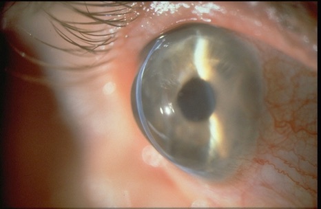
Rigid gas-permeable contact lenses, large-diameter or scleral lens designs that vault over the irregular inferior cornea, are frequently needed to achieve good functional vision once astigmatism becomes significantly irregular.
Corneal collagen cross-linking, discussed in more detail in its own dedicated article on this site for its broader use in ectatic disease, has been used to attempt to halt progression in pellucid marginal degeneration, though published outcomes are less extensive than for keratoconus given the disease’s relative rarity.
Surgical options for advanced or contact lens-intolerant disease are more technically demanding than for central keratoconus, given the eccentric, peripheral location of the thinning.
Crescentic lamellar keratoplasty and, in severe cases, large or eccentric penetrating keratoplasty have both been used, with generally more variable outcomes than standard central keratoplasty for keratoconus, in part because the peripheral, eccentric graft placement required is technically less forgiving and the resulting astigmatism can be harder to fully control even after a technically successful transplant.


Document what you see
Two smartphone imaging tools built for everyday clinic use — one for the slit lamp, one for the fundus.
From Choroida — the team behind this siteReferences
- Krachmer JH. Pellucid marginal corneal degeneration. Archives of Ophthalmology.
- Sridhar MS, Mahesh S, Bansal AK, et al. Pellucid marginal corneal degeneration. Ophthalmology.
- American Academy of Ophthalmology. Basic and Clinical Science Course, Section 8: External Disease and Cornea.
