
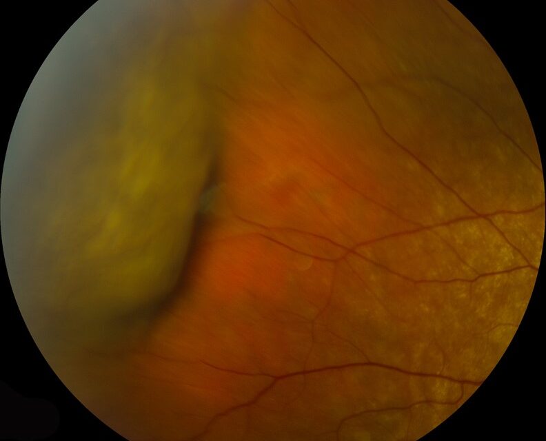
Choroidal detachment is a separation of the choroid from the overlying sclera, most often caused by abnormally low intraocular pressure (hypotony) that allows fluid to accumulate in the suprachoroidal space, or, less commonly, by suprachoroidal hemorrhage from a ruptured choroidal vessel.
It is a distinct entity from retinal detachment, though the two can coexist and can be confused with each other on a cursory exam, with important differences in management and urgency.
Mechanism
The suprachoroidal space is a potential space between the choroid and sclera that is normally kept closed by a small pressure gradient favoring fluid absorption.
When intraocular pressure drops significantly — after intraocular surgery, trauma with a wound leak, or an overfiltering glaucoma bleb — that gradient reverses, and transudative fluid accumulates in the suprachoroidal space, lifting the choroid away from the sclera.
Suprachoroidal hemorrhage follows a different, more acute mechanism: rupture of a long or short posterior ciliary artery, typically during or after intraocular surgery or from trauma, causing rapid blood accumulation in the same potential space.
Serous (Hypotony-Related) Choroidal Detachment
- Develops in the setting of low intraocular pressure, classically following intraocular surgery, a penetrating injury with a wound leak, or an overfiltering glaucoma drainage procedure
- Typically peripheral, often extending 360 degrees in more significant cases, with the classic “kissing choroidals” appearance when detachments from opposite sides of the eye meet in the middle
- Usually painless, and often discovered on routine postoperative exam rather than because of patient-reported symptoms
- Resolves as the underlying hypotony is corrected and normal pressure gradients are restored
Fundus Explorer Pro
Photograph the retinal findings described here with the phone already in your pocket — 22 D optics and built-in illumination in one handheld unit.
From Choroida — the team behind this siteSuprachoroidal Hemorrhage
- Presents acutely, often intraoperatively, with sudden pain, rising intraocular pressure (the opposite of hypotony), and shallowing of the anterior chamber
- A recognized, feared complication of intraocular surgery, particularly in eyes with risk factors such as high myopia, prior vitrectomy, elevated blood pressure, or anticoagulant use
- Can range from a limited, self-resolving hemorrhage to a massive, expulsive hemorrhage that pushes intraocular contents out through the surgical wound — a rare but catastrophic intraoperative event
Clinical Findings
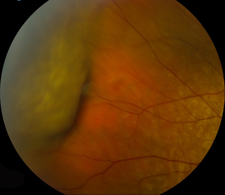
On fundus exam, a choroidal detachment appears as a smooth, dome-shaped, orange-brown elevation, distinctly different from the more corrugated, mobile appearance of a rhegmatogenous retinal detachment.
It does not extend to the ora serrata in the same way a retinal detachment can, because it is bound by the vortex vein ampullae.
B-scan ultrasonography is useful when the view is obscured by media opacity or hemorrhage, showing the characteristic smooth, dome-shaped elevation without the fixed folds typical of longstanding retinal detachment.
Differential Diagnosis
- Rhegmatogenous retinal detachment — corrugated, mobile, extends to the ora serrata, associated with a retinal break
- Choroidal tumor (melanoma, metastasis) — a discrete mass rather than a diffuse, dome-shaped elevation, with characteristic ultrasound reflectivity patterns distinguishing it from simple fluid or blood
- Posterior scleritis — inflammatory, painful, with characteristic scleral thickening on ultrasound (the “T-sign”)
Management
Serous choroidal detachment from hypotony is managed by identifying and correcting the underlying cause of low pressure — sealing a wound leak, addressing an overfiltering bleb, treating any coexisting inflammation — after which the detachment typically resolves spontaneously as normal pressure gradients are restored, without any direct intervention on the choroidal fluid itself.
Topical corticosteroids and cycloplegia are often used adjunctively to reduce inflammation and improve comfort while the underlying cause is addressed.
Suprachoroidal hemorrhage is managed based on severity: limited, self-resolving hemorrhage is observed, while extensive or “kissing” hemorrhage causing significant pain, elevated pressure, or apposition threatening the retina may require surgical drainage, typically performed after allowing the clot to liquefy over one to two weeks.
This makes drainage technically easier and reduces the risk of further bleeding compared with attempting evacuation acutely.
Prognosis
Serous choroidal detachment from correctable hypotony generally has a good prognosis once the underlying cause is addressed, with the choroid typically reattaching fully.
Suprachoroidal hemorrhage carries a more variable prognosis depending on its extent and whether it occurred intraoperatively or postoperatively, with large, expulsive hemorrhages carrying a guarded visual prognosis despite best surgical management.


Document what you see
Two smartphone imaging tools built for everyday clinic use — one for the slit lamp, one for the fundus.
From Choroida — the team behind this siteReferences
- Ho J, Regillo CD. Suprachoroidal hemorrhage. In: Ryan SJ, ed. Retina.
- Chu TG, Green RL. Suprachoroidal hemorrhage. Survey of Ophthalmology.
- American Academy of Ophthalmology. Basic and Clinical Science Course, Section 12: Retina and Vitreous.
- Bagheri N, Wajda B, eds. The Wills Eye Manual.
