
Case Study
A 32-year-old male presented with gradual blurring of vision and floaters in his left eye over several months. He denied pain or redness.
Family history was notable for renal tumors in a first-degree relative. Best-corrected visual acuity was 20/20 in the right eye and 20/40 in the left eye.
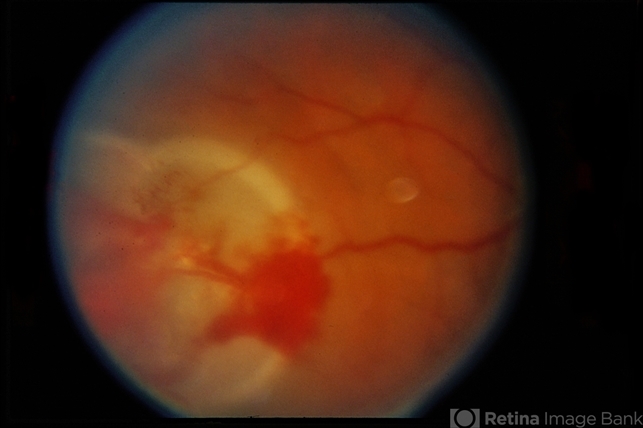
Fundus examination of the left eye revealed a well-circumscribed, reddish-orange vascular mass in the superotemporal peripheral retina, supplied by a dilated tortuous feeder arteriole and drained by an enlarged vein.
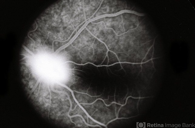
Surrounding lipid exudation and localized serous retinal detachment were noted. Fluorescein angiography showed early hyperfluorescence with intense late leakage.
A diagnosis of retinal capillary hemangioblastoma was made, and systemic evaluation was initiated to exclude von Hippel–Lindau disease.
Disease Entity
Retinal capillary hemangioblastoma (RCH), also known as retinal hemangioblastoma or retinal angioma, is a benign but potentially vision-threatening vascular tumor of the retina.
It is composed of proliferating capillary-sized blood vessels and stromal cells. These tumors may occur sporadically or as part of von Hippel–Lindau (VHL) disease, an autosomal dominant multisystem disorder.
Although histologically benign, retinal capillary hemangioblastomas can lead to significant visual morbidity due to exudation, retinal detachment, vitreous hemorrhage, and secondary glaucoma if left untreated.
Fundus Explorer Pro
Photograph the retinal findings described here with the phone already in your pocket — 22 D optics and built-in illumination in one handheld unit.
From Choroida — the team behind this sitePathophysiology
The tumor arises from abnormal proliferation of retinal capillaries, leading to the formation of a vascular mass with high permeability. Leakage of plasma lipids and fluid from these vessels results in:
-
Intraretinal and subretinal exudation
-
Macular edema
-
Serous or tractional retinal detachment
In VHL-associated cases, mutations in the VHL tumor suppressor gene lead to increased hypoxia-inducible factors (HIFs), promoting angiogenesis through upregulation of vascular endothelial growth factor (VEGF).
Etiology and Classification
Retinal capillary hemangioblastomas are classified into:
Sporadic
-
Usually solitary
-
Often diagnosed later in life
-
Less likely to have bilateral or multiple lesions
VHL-Associated
-
Frequently multiple and bilateral
-
Occur at a younger age
-
Associated with systemic tumors such as cerebellar hemangioblastomas, renal cell carcinoma, pheochromocytoma, and pancreatic cysts
Epidemiology
-
Typically diagnosed in the second to fourth decades of life
-
No strong gender predilection
-
Up to 60% of patients with retinal capillary hemangioblastoma have underlying VHL disease
-
Retinal involvement is often the earliest manifestation of VHL
Clinical Features
Symptoms depend on tumor size, location, and secondary complications.
Common presentations include:
-
Blurred or decreased vision
-
Floaters
-
Metamorphopsia
-
Visual field defects
Peripheral tumors may remain asymptomatic until significant exudation occurs, while juxtapapillary lesions can cause early visual loss.
Examination Findings
Fundus Examination
-
Round or oval, orange-red vascular mass
-
Prominent dilated feeder artery and draining vein
-
Surrounding lipid exudation
-
Localized or extensive serous retinal detachment
Imaging
-
Fluorescein angiography: Early hyperfluorescence with progressive leakage
-
OCT: Macular edema, subretinal fluid, and exudates
-
B-scan ultrasonography: Elevated retinal lesion with high internal reflectivity (for larger tumors)
Wide-field imaging is useful for detecting peripheral and multiple lesions.
Differential Diagnosis
-
Coats disease
-
Retinal vasoproliferative tumor
-
Choroidal hemangioma
-
Cavernous hemangioma of the retina
-
Inflammatory or neoplastic retinal masses
The presence of dilated feeder vessels is a key distinguishing feature.
Diagnosis
Diagnosis is primarily clinical, supported by angiographic findings. In all patients—especially young individuals or those with multiple or bilateral tumors—systemic evaluation for VHL disease is mandatory, including:
-
Neuroimaging
-
Abdominal imaging
-
Genetic testing when appropriate
Management
Treatment depends on tumor size, location, activity, and associated complications.
Observation
-
Small, inactive tumors without exudation
-
Close monitoring is essential
Local Therapies
-
Laser photocoagulation: Effective for small peripheral lesions
-
Cryotherapy: Preferred for larger peripheral tumors with subretinal fluid
-
Photodynamic therapy: Useful in selected cases
Adjunctive Treatments
-
Intravitreal anti-VEGF injections may reduce exudation but are rarely sufficient as monotherapy
Surgical Management
-
Pars plana vitrectomy may be required for:
-
Non-resolving retinal detachment
-
Vitreous hemorrhage
-
Epiretinal membrane formation
-
Prognosis
Visual prognosis depends on:
-
Tumor location (macular or juxtapapillary lesions have worse outcomes)
-
Degree of exudation and retinal detachment
-
Timeliness of treatment
Early detection and treatment can preserve good visual function, particularly in peripheral tumors.
Follow-Up and Systemic Implications
Patients require long-term ophthalmic follow-up due to the risk of:
-
Tumor recurrence
-
Development of new lesions
-
Progressive exudative complications
In VHL-associated cases, lifelong systemic surveillance is critical.


Document what you see
Two smartphone imaging tools built for everyday clinic use — one for the slit lamp, one for the fundus.
From Choroida — the team behind this siteReferences
-
Shields JA, Shields CL. Vascular tumors of the retina and optic disc. Ophthalmology.
-
Singh AD, Shields CL, Shields JA. Von Hippel–Lindau disease. Surv Ophthalmol.
-
American Academy of Ophthalmology. Basic and Clinical Science Course: Retina and Vitreous.
-
Yanoff M, Duker JS. Ophthalmology. 5th ed. Elsevier.
-
Tasman W, Jaeger EA. Duane’s Ophthalmology. Lippincott Williams & Wilkins.
Test yourself
A few questions straight from this article.
-
What is retinal capillary hemangioblastoma composed of?
Retinal capillary hemangioblastoma is a benign vascular tumour composed of proliferating capillary-sized blood vessels together with stromal cells. -
Which systemic disorder is retinal capillary hemangioblastoma classically associated with?
The tumour may be sporadic or part of von Hippel–Lindau disease, an autosomal dominant multisystem disorder, so systemic evaluation is mandatory. -
What proportion of patients with retinal capillary hemangioblastoma have underlying VHL disease?
Up to 60% of patients with retinal capillary hemangioblastoma have underlying von Hippel–Lindau disease, and retinal involvement is often the earliest manifestation of VHL. -
How does VHL gene mutation drive tumour growth in retinal capillary hemangioblastoma?
Loss of the VHL tumour suppressor raises hypoxia-inducible factors, which promote angiogenesis through upregulation of vascular endothelial growth factor. -
Which fundus finding is the key clue to retinal capillary hemangioblastoma?
The tumour appears as a round or oval orange-red vascular mass with a prominent dilated feeder artery and draining vein; these feeder vessels are the key distinguishing feature. -
How do VHL-associated retinal capillary hemangioblastomas differ from sporadic ones?
VHL-associated tumours are frequently multiple and bilateral and occur at a younger age, whereas sporadic lesions are usually solitary and diagnosed later in life. -
Which fluorescein angiography pattern is seen in retinal capillary hemangioblastoma?
FA shows early hyperfluorescence of the tumour with progressive, intense late leakage, reflecting the high permeability of the proliferating vessels. -
Which treatment is preferred for a large peripheral retinal capillary hemangioblastoma with subretinal fluid?
Cryotherapy is preferred for larger peripheral tumours with subretinal fluid, while laser photocoagulation suits small peripheral lesions. -
What role does intravitreal anti-VEGF have in retinal capillary hemangioblastoma?
Anti-VEGF injections are adjunctive: they may reduce exudation but are rarely sufficient as monotherapy. -
Which tumour location carries the worst visual prognosis in retinal capillary hemangioblastoma?
Visual prognosis depends on location, and macular or juxtapapillary lesions have worse outcomes; juxtapapillary tumours can cause early visual loss.
