
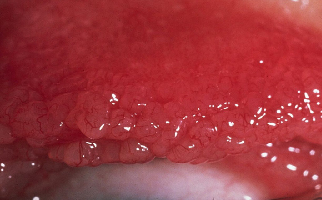
Giant papillary conjunctivitis (GPC) is a chronic inflammatory reaction of the upper tarsal conjunctiva driven by mechanical irritation and, frequently, an immune response to protein deposits on a foreign surface — most commonly a contact lens, though ocular prostheses and exposed sutures can trigger the same process.
It is one of the more common reasons a contact lens wearer ends up needing to stop lens wear, at least temporarily, and recognizing it early changes the entire trajectory of management.
Pathogenesis
Two mechanisms act together: mechanical trauma from the lens edge or surface repeatedly rubbing against the upper tarsal conjunctiva with every blink, and an immune-mediated hypersensitivity response to protein and lipid deposits that accumulate on the lens surface over time.
This combination — mechanical plus immunologic — is why GPC develops gradually over months of lens wear rather than acutely, and why simply cleaning the lens more thoroughly is rarely sufficient once established papillae have formed.
Once papillae have formed, they represent a genuine structural change in the conjunctival tissue rather than a purely reversible inflammatory state, which is part of why treatment focuses as much on removing the ongoing trigger as on resolving the current inflammation — simply treating the inflammation while lens wear continues tends to produce only partial, temporary improvement.
Risk Factors
- Soft contact lens wear, particularly with poor lens hygiene or infrequent replacement
- Extended or overnight lens wear
- Ocular prosthesis wear
- Exposed monofilament sutures following intraocular or corneal surgery
- Filtering blebs following glaucoma surgery, in some reported cases
- A personal history of atopy, which appears to increase susceptibility
All-fit Slit-Lamp Adapter
Record and share exactly what you see at the slit lamp. One adapter fits any slit lamp or surgical microscope — and any smartphone.
From Choroida — the team behind this siteClinical Presentation
Patients describe itching (often the dominant symptom), mucous discharge, contact lens intolerance with increasing awareness of the lens on the eye, and blurred vision from mucus coating the lens surface.
Symptoms characteristically worsen with continued lens wear and improve, at least partially, with any period of discontinuation, which is itself a useful diagnostic and prognostic clue.
Because the earliest symptoms are often subtle and easy to attribute to ordinary lens dryness or fatigue, many patients have been symptomatic for weeks to months, gradually reducing their own wearing time to cope, before the underlying papillary change is formally identified on exam.
Exam Findings and Staging
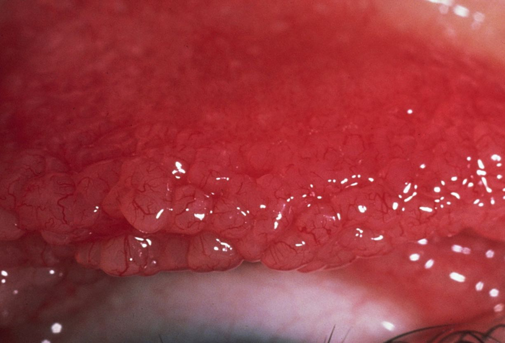
- Giant papillae (typically defined as greater than 0.3 mm, sometimes reaching a cobblestone appearance) on the superior tarsal conjunctiva, best seen with lid eversion
- Milder, earlier disease shows a fine papillary reaction rather than the fully developed giant papillae of established disease
- Mucous discharge, sometimes stringy, adherent to the lens or lid margin
- Mild conjunctival injection
- Reduced lens tolerance and wearing time as a functional marker of severity
Because the earliest fine papillary changes are easy to miss without deliberately everting the upper lid, this maneuver should be a routine part of the exam in any contact lens wearer reporting new or worsening lens intolerance, even before giant papillae are suspected.
Differential Diagnosis
- Vernal keratoconjunctivitis — giant papillae as well, but typically with more severe symptoms, seasonal pattern, Horner-Trantas dots at the limbus, and onset independent of contact lens wear
- Superior limbic keratoconjunctivitis — superior bulbar and limbal involvement rather than tarsal papillae, often associated with thyroid disease
- Bacterial or allergic conjunctivitis — different discharge pattern and time course, without the giant papillary reaction
- Floppy eyelid syndrome — mechanically distinct, related to lid laxity rather than a lens or foreign body trigger
The history of contact lens or prosthesis wear, together with the characteristic giant papillae confined to the upper tarsal conjunctiva, is usually sufficient to separate GPC from these alternatives without further testing.
Management
Discontinuing or reducing contact lens wear is the single most effective intervention and is often necessary at least temporarily, even though it is rarely the answer patients want to hear.
When lens wear resumes, switching to a different lens material (particularly daily disposable lenses, which eliminate the protein buildup that accumulates on reusable lenses over time), more frequent lens replacement, and more rigorous cleaning regimens all reduce the antigenic and mechanical burden driving the reaction.
Topical mast cell stabilizers or combination antihistamine/mast cell stabilizer drops address the allergic component and are commonly used both during active disease and prophylactically once lens wear resumes.
Short courses of topical corticosteroids can be used for more severe or refractory flares.
For GPC driven by exposed sutures or a prosthesis, removing or adjusting the offending suture or refitting the prosthesis addresses the mechanical trigger directly.
Patient education about the underlying mechanism is genuinely useful here, since understanding that the papillae reflect a real, cumulative reaction to the lens surface — rather than a simple infection that antibiotics might clear — helps patients accept why a period of reduced or discontinued lens wear is necessary even when the eye does not look dramatically inflamed.
Prognosis
GPC generally resolves with removal of the inciting trigger, though established giant papillae can take weeks to months to fully regress even after the trigger is removed.
Some patients are left with a degree of chronic papillary change that limits future lens tolerance.
With appropriate lens modification and hygiene changes, many patients can successfully resume contact lens wear once the acute inflammation has settled, though a subset find they tolerate lens wear best only with the ongoing, deliberate combination of daily disposables and ongoing prophylactic anti-allergy drops.


Document what you see
Two smartphone imaging tools built for everyday clinic use — one for the slit lamp, one for the fundus.
From Choroida — the team behind this siteReferences
- Allansmith MR, Korb DR, Greiner JV, et al. Giant papillary conjunctivitis in contact lens wearers. American Journal of Ophthalmology.
- Elhers WH, Donshik PC. Giant papillary conjunctivitis. Current Opinion in Allergy and Clinical Immunology.
- American Academy of Ophthalmology. Basic and Clinical Science Course, Section 8: External Disease and Cornea.
