
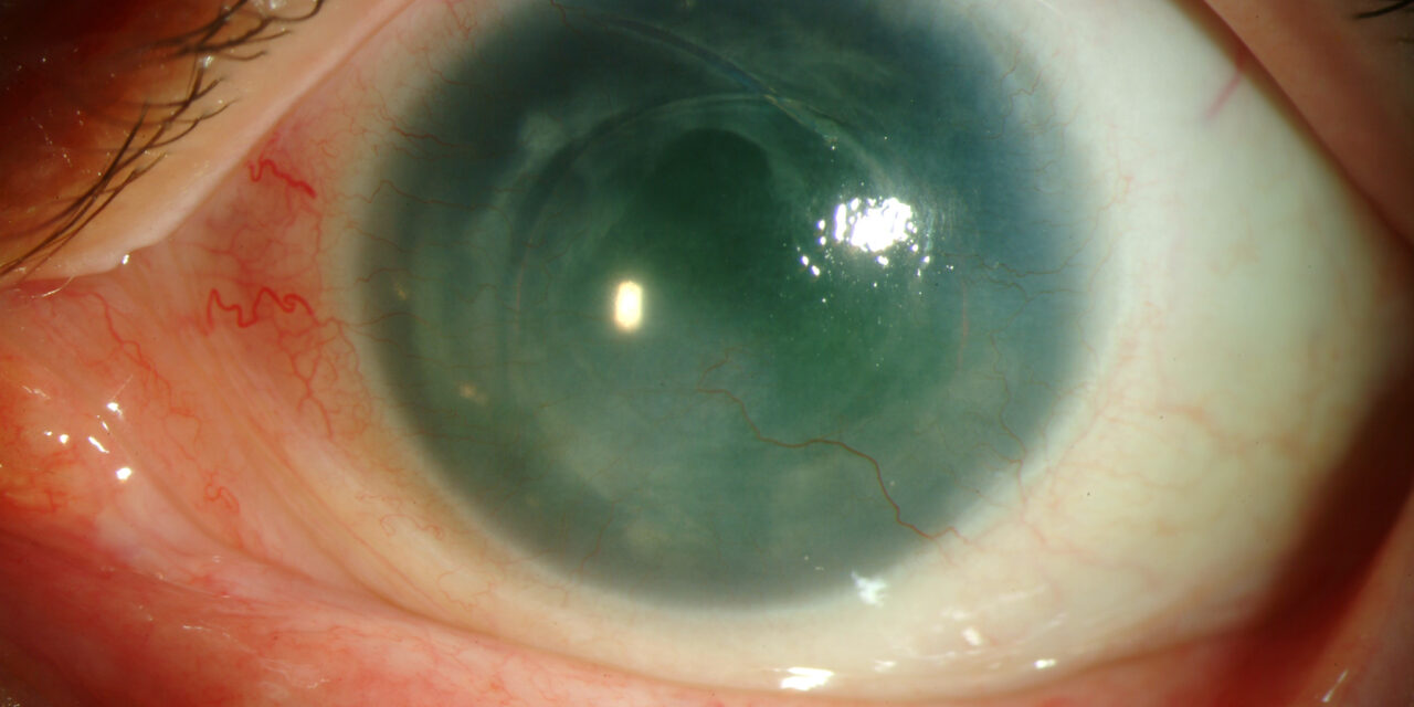
Aniridia-related keratopathy (ARK) is a progressive corneal surface disease that develops in a large majority of patients with congenital aniridia over their lifetime, and for many of these patients it — not the absent iris itself — ends up being the dominant threat to vision.
The near-absence of the iris that defines aniridia is only the most visually obvious part of a much broader anterior segment developmental disorder, and ARK is one of its most consequential downstream consequences.
Pathogenesis
Congenital aniridia is caused by heterozygous mutations or deletions involving PAX6, a master regulatory gene essential for normal eye development, whose effects extend well beyond the iris to the cornea, lens, macula, and optic nerve.
Limbal stem cell deficiency is now understood to be the central mechanism driving ARK: the limbus, which normally houses the stem cell population responsible for continuously regenerating corneal epithelium, is structurally and functionally abnormal in aniridia, leading to progressive failure of normal corneal epithelial maintenance over years.
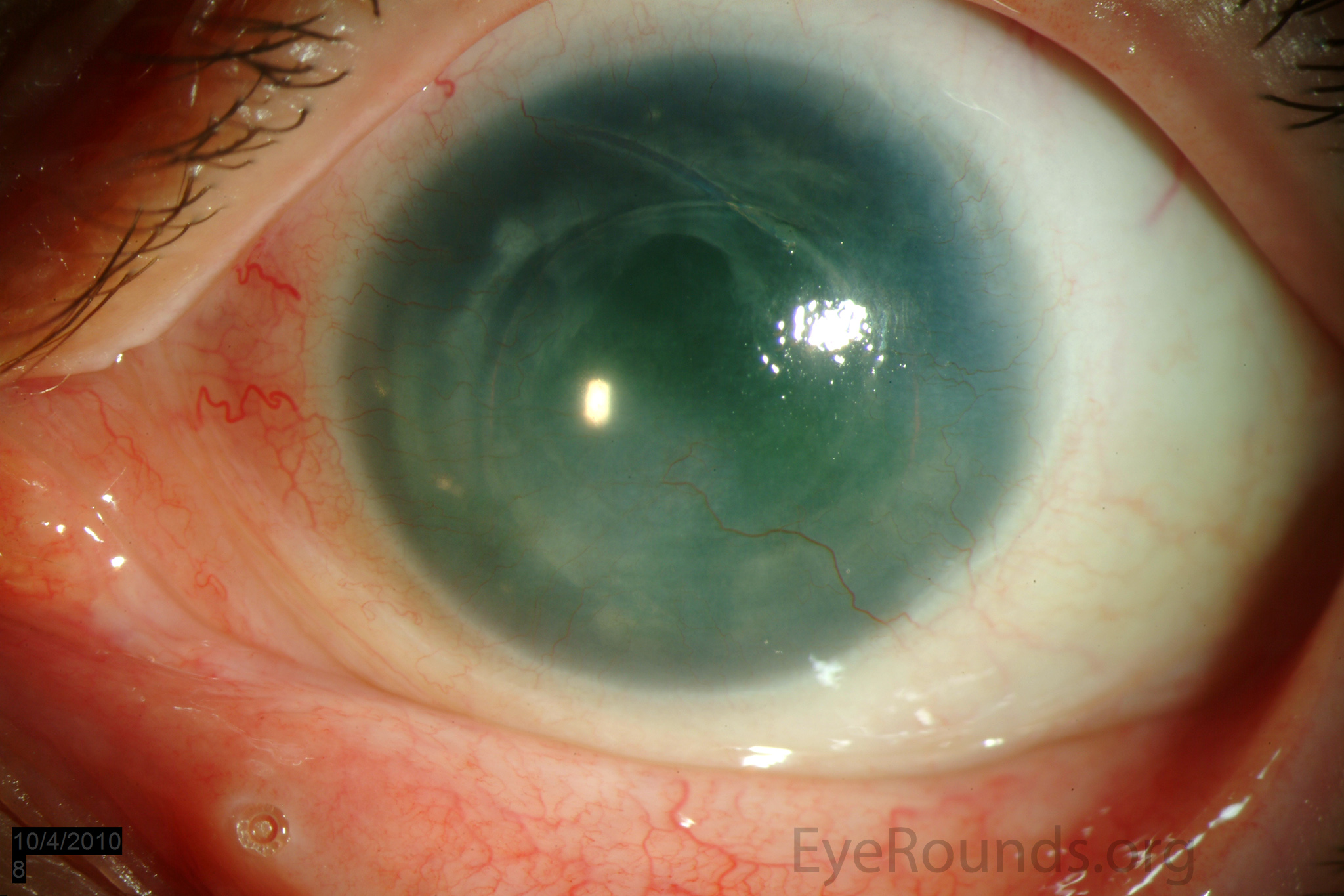
As limbal stem cell function declines, conjunctival epithelium — which is not meant to cover the cornea — gradually migrates centrally across the corneal surface, bringing goblet cells, subepithelial fibrovascular tissue, and chronic inflammation with it, in a process called conjunctivalization that is the hallmark of ARK.
Clinical Staging
ARK typically progresses through recognizable stages, beginning with mild peripheral corneal haze and subtle limbal changes, advancing through progressive conjunctivalization and pannus formation encroaching on the visual axis, and culminating in severe cases with dense central corneal scarring, vascularization, and significant vision loss.
The pace of progression varies considerably between patients and even between the two eyes of the same patient, though the underlying trajectory — from limbal compromise to central corneal involvement — is broadly consistent.
All-fit Slit-Lamp Adapter
Record and share exactly what you see at the slit lamp. One adapter fits any slit lamp or surgical microscope — and any smartphone.
From Choroida — the team behind this siteClinical Findings
- Peripheral corneal haze and subepithelial fibrosis, often the earliest sign
- Progressive pannus — fibrovascular tissue advancing from the limbus toward the central cornea
- Irregular, thickened, poorly wetting corneal epithelium
- Recurrent or persistent epithelial defects in more advanced disease
- Dry eye, which is common in aniridia independent of ARK and compounds the surface disease
- Reduced corneal sensation in some patients, adding a neurotrophic component to an already compromised surface
Compounding Factors
Several other features common in aniridia make ARK worse or harder to manage once it develops: chronic photophobia from the absent iris often leads patients to rub their eyes excessively, which mechanically stresses an already fragile limbal stem cell population.
Aniridia-associated glaucoma, present in a substantial proportion of patients, requires topical medications whose preservatives can further damage an already compromised ocular surface, and cataract surgery, needed by many aniridic patients at a young age, carries its own additional risk of further limbal stem cell injury from surgical trauma.
Differential Diagnosis of Progressive Corneal Conjunctivalization
- Other causes of limbal stem cell deficiency — chemical burns, Stevens-Johnson syndrome, ocular cicatricial pemphigoid, contact lens overwear — distinguished from ARK by the absence of aniridia and a different inciting history
- Chronic ocular surface disease from severe dry eye alone, without true limbal stem cell loss
- Pterygium — a localized, wedge-shaped fibrovascular growth rather than the circumferential, progressive process typical of ARK
Management
Aggressive preservative-free lubrication is foundational and should be started proactively, not reactively, given how predictably ARK develops in this population, aiming to reduce mechanical and inflammatory stress on an already vulnerable limbus.
Minimizing preserved topical medications — relevant given how often these patients also need glaucoma drops — and addressing photophobia (tinted lenses, environmental modification) to reduce the eye rubbing that mechanically accelerates limbal damage are both practical, proactive measures.
For more advanced disease, limbal stem cell transplantation (from a living related donor, cadaveric tissue, or, increasingly, the patient’s own healthy limbal tissue when only one eye is affected) can restore a functional corneal epithelium, though outcomes are limited by the same underlying ocular surface inflammation and dryness that caused the deficiency, and results are generally less durable than limbal transplantation for other causes.
Amniotic membrane transplantation has a role in managing acute epithelial defects and reducing surface inflammation, and, in end-stage disease with extensive scarring, keratoprosthesis is sometimes considered, though the compromised surface makes these eyes a challenging substrate even for an artificial cornea, and outcomes in this population tend to be more variable than in eyes with a healthier baseline ocular surface.
Why Early, Proactive Management Matters
Because ARK is a slow, progressive, largely predictable consequence of the underlying limbal stem cell deficiency in aniridia, the most effective strategy is proactive surface protection started early — well before significant conjunctivalization develops — rather than waiting to intervene once vision is already threatened.
This shifts the practical focus of aniridia eye care substantially toward ocular surface maintenance, alongside the glaucoma and cataract management that more commonly dominate discussion of the condition.
Families of children with newly diagnosed aniridia benefit from understanding, early on, that ARK is not a rare or unlikely complication but an expected part of the disease’s natural history for most patients, so that lubrication and photophobia management become an established habit rather than an afterthought introduced only once surface disease has already progressed.


Document what you see
Two smartphone imaging tools built for everyday clinic use — one for the slit lamp, one for the fundus.
From Choroida — the team behind this siteReferences
- Lee H, Khan R, O’Keefe M. Aniridia: current pathology and management. Acta Ophthalmologica.
- Nishida K, Kinoshita S, Ohashi Y, et al. Ocular surface abnormalities in aniridia. American Journal of Ophthalmology.
- Lagali N, Wowra B, Fries FN, et al. Early phenotypic features of aniridia-associated keratopathy and association with PAX6 coding mutations. Ocular Surface.
- American Academy of Ophthalmology. Basic and Clinical Science Course, Section 8: External Disease and Cornea.
