
Cat eye syndrome is a rare chromosomal disorder caused by partial tetrasomy of chromosome 22 (an extra, small supernumerary marker chromosome containing duplicated material from the 22q11.2 region).
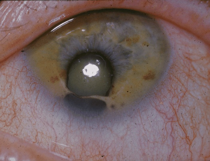
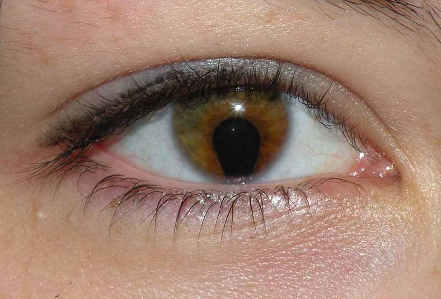
It takes its name directly from its most recognizable ocular feature: bilateral iris and/or chorioretinal coloboma, combined with variably present preauricular skin tags and pits, giving some affected individuals a distinctive vertically slit-like pupil reminiscent of a cat’s eye.
The syndrome illustrates well how a single, memorable physical sign can anchor recognition of a much broader multisystem condition.
Genetics
The extra marker chromosome in cat eye syndrome contains a duplicated segment of the proximal long arm of chromosome 22, distinguishing it genetically from 22q11.2 deletion syndrome (DiGeorge/velocardiofacial syndrome), which involves a deletion rather than a duplication in a related but not identical region.
Despite the similar-sounding chromosomal location, the two conditions are genetically and clinically distinct entities, and mixing them up in a differential diagnosis discussion is a common error worth actively avoiding.
Most cases arise sporadically, though the marker chromosome can occasionally be inherited from a mosaic or balanced-carrier parent, which is why genetic counseling. Relevant parental chromosomal testing is part of a complete workup once the diagnosis is confirmed in a child.
The degree of tetrasomy — how much duplicated material the marker chromosome actually carries — varies between patients and appears to correlate at least loosely with phenotypic severity, which is one reason the clinical presentation of cat eye syndrome spans such a wide range, from mildly affected individuals detected almost incidentally to those with severe, multi-organ involvement.
Ocular Findings
- Iris coloboma — typically inferonasal, ranging from a subtle notch to a full-thickness keyhole defect
- Chorioretinal coloboma, which can affect the optic nerve and macula depending on extent, with corresponding visual field or acuity impact
- Microphthalmia in some affected eyes
- Strabismus
- Duane retraction syndrome, reported in some cases
Not every affected individual has the classic cat’s-eye pupil appearance; coloboma severity varies widely, and some patients have only mild or unilateral involvement.
This means the syndrome should still be considered even when the ocular finding is subtle, provided the systemic features described below are present.
A full dilated fundus exam is worthwhile in any suspected case rather than relying on external inspection of the iris alone, since chorioretinal coloboma is not visible without dilation.
All-fit Slit-Lamp Adapter
Record and share exactly what you see at the slit lamp. One adapter fits any slit lamp or surgical microscope — and any smartphone.
From Choroida — the team behind this siteSystemic Features
- Preauricular skin tags and pits — often the most consistently present external finding, sometimes even more reliably present than the coloboma itself
- Anal atresia or other anorectal malformations
- Congenital heart defects, particularly total anomalous pulmonary venous return and other structural anomalies
- Renal anomalies
- Variable intellectual disability, ranging from normal cognitive function to significant impairment
- Mild dysmorphic facial features in some patients
The combination of preauricular tags/pits, anal atresia, and coloboma or other ocular anomaly should prompt specific consideration of cat eye syndrome and genetic testing, because the syndrome’s variable expressivity means no single feature is reliably present in every case.
Differential Diagnosis
- CHARGE syndrome — coloboma is shared, but the broader feature set (choanal atresia, characteristic ear anomalies, growth deficiency) differs, and the genetics are distinct (typically CHD7 mutations rather than a chromosome 22 marker)
- 22q11.2 deletion syndrome — genetically distinct (deletion, not duplication), with a different, though partially overlapping, phenotype (cardiac defects, immune dysfunction, palatal anomalies)
- Isolated coloboma without a syndromic association — the ocular finding alone, without the systemic features characteristic of cat eye syndrome
- Goldenhar syndrome (oculo-auriculo-vertebral spectrum) — also features preauricular tags and ocular anomalies (typically epibulbar dermoid rather than coloboma), but with distinct vertebral and mandibular features
Diagnostic Evaluation
Chromosomal microarray or specific FISH testing for the chromosome 22 marker confirms the diagnosis once clinically suspected.
A full systemic evaluation — echocardiogram, renal ultrasound, and assessment for anorectal anomalies — is essential once the diagnosis is made or strongly suspected, given the range of associated malformations that can be present and require their own dedicated management.
Because anorectal malformations may not be immediately obvious on casual newborn exam, a specific, deliberate assessment for anal atresia and related anomalies is worthwhile in any newborn with the combination of preauricular tags and ocular coloboma, rather than assuming a normal-appearing perineal exam has adequately excluded this possibility.
Management
There is no treatment for the underlying chromosomal abnormality; management is directed at the individual manifestations.
Surgical correction of significant coloboma-related complications where feasible, cardiac and renal management as indicated by the specific anomalies present, and developmental support tailored to the child’s cognitive profile are the mainstays of care.
Ophthalmic follow-up focuses on monitoring visual development, treating any amblyopia from coloboma-related visual impairment or strabismus, and managing the anatomic risks associated with coloboma, including an elevated risk of retinal detachment in eyes with chorioretinal involvement.
Given the number of organ systems potentially involved, coordinated multidisciplinary care — cardiology, nephrology, genetics, and developmental pediatrics working alongside ophthalmology — is central to comprehensive long-term management, with the specific combination of specialists involved tailored to each child’s particular pattern of findings and reassessed periodically as the child grows and new developmental or medical needs emerge.


Document what you see
Two smartphone imaging tools built for everyday clinic use — one for the slit lamp, one for the fundus.
From Choroida — the team behind this siteReferences
- Schinzel A, Schmid W, Fraccaro M, et al. The “cat eye syndrome”: dicentric small marker chromosome probably derived from a No.22 (tetrasomy 22pter-q11) associated with a characteristic phenotype. Human Genetics.
- McTaggart KE, Budarf ML, Driscoll DA, et al. Cat eye syndrome chromosome breakpoint clustering: identification of two intervals also associated with 22q11 deletion syndrome breakpoints. Cytogenetics and Cell Genetics.
- American Academy of Ophthalmology. Basic and Clinical Science Course, Section 6: Pediatric Ophthalmology and Strabismus.
