
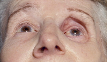
Enophthalmos — posterior displacement of the globe within the orbit — is the mirror image of proptosis.
Like proptosis, it is a physical sign rather than a diagnosis, produced whenever the orbit loses volume, gains space, or has its normal soft-tissue support disrupted.
It is generally subtler and easier to miss on casual inspection than proptosis, which often means the diagnosis is delayed until the asymmetry becomes pronounced or a family member points it out.
Because the underlying process — bony expansion, fat loss, or soft-tissue contraction — can be silent for a long time before the resulting globe displacement becomes visually obvious, a systematic approach to evaluation matters more here than it might for a more dramatically presenting sign.
Mechanisms
Enophthalmos develops through a few distinct routes: an enlarged bony orbit (from fracture) that gives orbital contents more room to settle backward, loss of orbital fat volume (from atrophy, trauma, or surgery), a defect in the orbital walls allowing soft tissue to herniate into an adjacent sinus, or, less commonly, a slow-growing sclerosing process that contracts orbital tissue over time.
The common thread across all these mechanisms is a change in the volume relationship between the bony orbit and its contents, pushing the balance toward the globe sitting further back than normal.
Common Causes
- Orbital blowout fracture — the most common acute cause, from expansion of the orbital volume when the thin orbital floor or medial wall fractures outward
- Silent sinus syndrome — chronic maxillary sinus atelectasis causing gradual, painless orbital floor collapse and enophthalmos, without any preceding trauma
- Fat atrophy — age-related, or following significant weight loss, chronic steroid use, or prior orbital surgery or radiation
- Sclerosing orbital metastasis — classically from scirrhous breast carcinoma, one of the few malignancies that causes enophthalmos rather than the proptosis typical of most orbital tumors
- Linear scleroderma (en coup de sabre) — localized soft-tissue atrophy involving the orbit
- Congenital microphthalmia or anophthalmia — a structurally small or absent globe producing an enophthalmic appearance from birth
All-fit Slit-Lamp Adapter
Record and share exactly what you see at the slit lamp. One adapter fits any slit lamp or surgical microscope — and any smartphone.
From Choroida — the team behind this siteClinical Evaluation
Exophthalmometry, the same tool used to quantify proptosis, works equally well to document enophthalmos and, as with proptosis, its real value is in tracking change over serial visits rather than a single measurement in isolation.
Comparing the two eyes side by side, checking the position of the superior sulcus (often deepened in enophthalmos as orbital fat volume is lost), and a full motility exam to check for any associated restriction complete the basic bedside assessment.
A history focused on trauma — even remote, since silent sinus syndrome and slow post-fracture changes can present well after the inciting event — unexplained weight loss, and known malignancy all matter here.
The tempo of onset — sudden versus gradual — helps direct the workup before imaging is even obtained, in much the same way it does for other orbital signs discussed elsewhere on this site.
Silent Sinus Syndrome: A Distinct Pattern Worth Knowing
Silent sinus syndrome deserves specific mention because it is easy to miss: patients present with gradually progressive, painless enophthalmos and a deepened superior sulcus, without any history of trauma, and often without significant sinus symptoms, despite chronic maxillary sinus obstruction and progressive orbital floor collapse being the underlying cause.
Recognizing the pattern — an otherwise unexplained, slowly progressive enophthalmos in an adult — should prompt sinus and orbital imaging even in the absence of a clear inciting event, since patients themselves are often unaware of any underlying sinus problem and would not think to mention it.
Differential Diagnosis of Apparent Enophthalmos
- True enophthalmos — actual posterior globe displacement, confirmed by exophthalmometry
- Contralateral proptosis — the “normal” eye is simply being compared to a proptotic fellow eye, making the normal side look falsely enophthalmic
- Ptosis with a deep superior sulcus — can mimic enophthalmos by narrowing the palpebral fissure and creating a similar visual impression without true posterior globe displacement
- Microphthalmia or phthisis bulbi — a genuinely small globe rather than a normal globe displaced posteriorly
Diagnostic Evaluation
CT of the orbits and sinuses is the primary imaging study, because it characterizes the bony orbital walls, any fracture or sinus disease, and orbital fat volume directly, and is the study of choice when trauma or silent sinus syndrome is suspected.
MRI adds value when a soft-tissue infiltrative or sclerosing process (such as metastatic breast carcinoma or scleroderma) is being considered, given its superior soft-tissue contrast compared to CT, particularly when the clinical picture and CT findings together do not fully explain the degree of enophthalmos present.
Management
Management addresses the underlying cause: orbital floor reconstruction for a blowout fracture with significant volume expansion, functional endoscopic sinus surgery combined with orbital floor repair for silent sinus syndrome, and, where a malignancy is identified, oncologic management with orbital involvement addressed as part of the broader treatment plan.
Cosmetic orbital volume augmentation (fillers or implants) is occasionally used for isolated fat atrophy once any underlying systemic or structural cause has been excluded or treated.
Pursuing cosmetic correction before completing this exclusion is generally discouraged, since a volume-restoring procedure performed on an eye with an unrecognized, ongoing underlying process (an undiagnosed malignancy or progressive sinus disease, for example) risks masking a condition that still requires its own specific diagnosis and treatment.


Document what you see
Two smartphone imaging tools built for everyday clinic use — one for the slit lamp, one for the fundus.
From Choroida — the team behind this siteReferences
- Soparkar CN, Patrinely JR, Cuaycong MJ, et al. The silent sinus syndrome. Ophthalmology.
- Rootman J, ed. Diseases of the Orbit: A Multidisciplinary Approach.
- American Academy of Ophthalmology. Basic and Clinical Science Course, Section 7: Orbit, Eyelids, and Lacrimal System.
- Bagheri N, Wajda B, eds. The Wills Eye Manual.
