
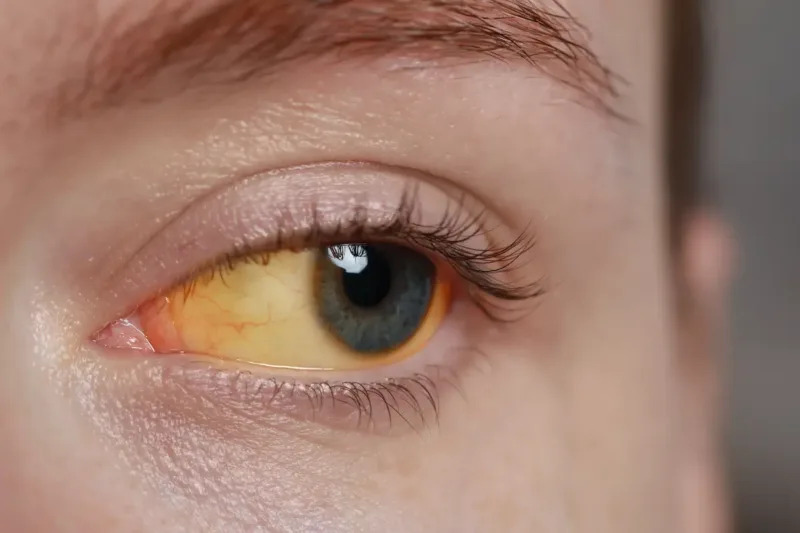
Scleral icterus — yellow discoloration of the sclera from bilirubin deposition — is often the first visible sign of hyperbilirubinemia, appearing before skin jaundice becomes obvious to the eye. The sclera’s high elastin content binds bilirubin readily, which is why the eyes can look yellow while the skin still looks close to normal, particularly in patients with darker skin tones where cutaneous jaundice is harder to appreciate.
Despite the name, the yellow color actually sits mostly in the conjunctiva overlying the sclera rather than the sclera itself, since conjunctival tissue has a rich vascular supply that delivers circulating bilirubin to the site, while the avascular sclera stains more slowly. The distinction rarely changes clinical management, but it explains why the discoloration can fluctuate somewhat faster than a true structural change in the sclera would.
Physiology of Bilirubin and the Eye
Bilirubin is a breakdown product of heme, produced mainly from senescent red blood cells and processed through unconjugated (indirect) and conjugated (direct) forms by the liver before excretion in bile. Scleral icterus typically becomes visible once total serum bilirubin rises above roughly 2 to 3 mg/dL, well before the level (usually cited around 3 mg/dL and higher) at which skin jaundice is reliably noticeable, making the eye a genuinely earlier clinical signal.
Because the finding reflects total bilirubin rather than distinguishing conjugated from unconjugated forms, scleral icterus by itself does not localize the cause — that requires laboratory fractionation and, usually, additional testing.
Causes
- Prehepatic — hemolysis (autoimmune, hereditary spherocytosis, glucose-6-phosphate dehydrogenase deficiency), ineffective erythropoiesis
- Hepatic — viral hepatitis, alcoholic or non-alcoholic liver disease, cirrhosis, Gilbert syndrome, drug-induced liver injury
- Posthepatic (obstructive) — choledocholithiasis, pancreatic or biliary malignancy, primary sclerosing cholangitis, biliary strictures
The pattern of associated findings helps narrow the category before labs come back: pale skin and dark urine with normal stool color suggest hemolysis, while pruritus, pale stool, and dark urine point toward obstruction, and stigmata of chronic liver disease (spider angiomata, palmar erythema, ascites) suggest a hepatic process.
All-fit Slit-Lamp Adapter
Record and share exactly what you see at the slit lamp. One adapter fits any slit lamp or surgical microscope — and any smartphone.
From Choroida — the team behind this siteExam Findings and Associated Signs
- Yellow discoloration most visible in natural light, examined against the white background of the sclera
- Conjunctival vessels may appear to run over, rather than under, the yellow discoloration, consistent with its predominantly conjunctival location
- Kayser-Fleischer rings (a separate, copper-related finding) should not be confused with icterus — they are a golden-brown limbal ring rather than diffuse yellowing
- Xanthelasma may coexist in patients with chronic cholestatic liver disease and lipid abnormalities, though it is a distinct finding from icterus itself
Differential Diagnosis of Yellow-Appearing Sclera
- True scleral icterus from hyperbilirubinemia
- Pinguecula — localized, yellow, raised, nasal or temporal, not diffuse
- Senile scleral hyaline plaque — localized gray-yellow patch anterior to the horizontal rectus insertions in older adults
- Carotenemia — orange-yellow skin discoloration from excess carotene intake, but the sclera itself stays white, which is the key distinguishing feature
Carotenemia is worth specifically excluding, since it can mimic jaundice on the skin quite convincingly but spares the sclera — a genuinely useful bedside distinction that avoids an unnecessary liver workup.
Evaluation
A patient found to have scleral icterus needs a total and direct (conjugated) bilirubin, liver enzymes (AST, ALT, alkaline phosphatase, GGT), and a complete blood count to screen for hemolysis. The pattern of these results — hepatocellular versus cholestatic versus a predominantly unconjugated hyperbilirubinemia — directs further imaging, typically abdominal ultrasound as a first step when biliary obstruction is suspected.
History should cover alcohol use, medications and supplements, viral hepatitis risk factors, recent travel, and family history, since Gilbert syndrome — a common, benign cause of mild unconjugated hyperbilirubinemia — is frequently uncovered incidentally in otherwise healthy people with intermittent scleral icterus during minor illness or fasting.
Clinical Significance for the Eye Care Provider
An ophthalmologist or optometrist noticing scleral icterus during a routine exam is often the first person to flag a new or worsening liver problem, particularly in patients who do not see a primary care provider regularly. This makes it one of the more consequential incidental findings in general eye examination, worth documenting and referring even when it is not the reason for the visit.
The finding itself requires no ocular treatment; management is entirely directed at the underlying systemic cause once identified.


Document what you see
Two smartphone imaging tools built for everyday clinic use — one for the slit lamp, one for the fundus.
From Choroida — the team behind this siteReferences
- Roche SP, Kobos R. Jaundice in the adult patient. American Family Physician.
- Fargo MV, Grogan SP, Saguil A. Evaluation of jaundice in adults. American Family Physician.
- Winger J, Michelfelder A. Diagnostic approach to the patient with jaundice. Primary Care.
- American Academy of Ophthalmology. Basic and Clinical Science Course, Section 8: External Disease and Cornea.
