
CASE REPORT
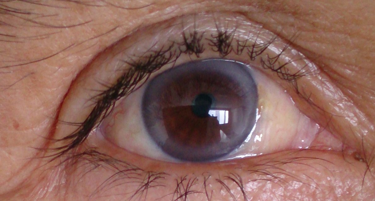
Case of Arcus Senilis a 55-year-old male patient presented with complaints of blurred vision in both eyes for 2 years. The patient was a known case of nephrotic syndrome with dyslipidemia for which he was on diuretics and lipid-lowering agents for 3 years.

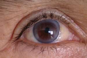
On examination, his visual acuity was 6/9 in both eyes with cloudy cornea and arcus juvenilis. Fundus examination was within normal limits.
On systemic workup, his lipid profile was deranged with increased serum total cholesterol, very low-density lipoprotein, low-density lipoprotein, and triglyceride.
The serum high-density lipoprotein was decreased. Renal function test revealed elevated serum creatinine with significant proteinuria.
An ocular manifestation of dense deposit disease is characterized by retinal drusen, pigmentary atrophy, choroidal neovascular membrane, and atypical serous retinopathy. The case was documented as Arcus Senilis.
DISEASE of Arcus Senilis
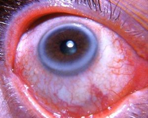
Also known as gerontoxon, arcus lipoides, arcus cornea, or corneal arcus, is a deposition of lipid in the peripheral corneal stroma. It is the most common peripheral corneal opacity.
Frequently it occurs with hyperlipidemia, especially in elderly individuals, and may be associated with dyslipidemia in younger patients (termed arcus juvenilis).
MANAGEMENT of Arcus Senilis
Patients can be followed at yearly intervals as would be expected for follow-up in a patient with a normal eye exam. There is no specific management necessary.
Prominent Arcus Senilis is often associated with decreased clarity on surgical view, as are other stromal opacities.
HOW TO TAKE Slit-lamp exam IMAGES BY A SMARTPHONE?
Smartphone slit-lamp photography is the new advancement in the field of science and technology in which the photographs of the desired slit-lamp finding can be taken with smartphones by using the slit-lamp adaptors.

Know more about Crocodile Shagreen, Christmas tree cataract, Bietti Crystalline Dystrophy, Branch Retinal Artery Occlusion
REFERENCES
- Kanski, J. J. & Bowling, B. Clinical Ophthalmology: A Systematic Approach. (Elsevier Health Sciences, 2011).
- Hashemi, H., Khabazkhoob, M., Emamian, M. H., Shariati, M. & Fotouhi, A. A population-based study of corneal arcus and its risk factors in Iran. Ophthalmic Epidemiol. 21, 339–344 (2014).
- Ang, M. et al. Corneal Arcus is a Sign of Cardiovascular Disease, Even in Low-Risk Persons. Am. J. Ophthalmol. 152, 864–871.e1 (2011).
- Patterson, L. Arcus senilis. Am. J. Forensic Med. Pathol. 3, 115–118 (1982).
- Raj, K. M., Reddy, P. A. S. & Kumar, V. C. Significance of corneal arcus. J. Pharm. Bioallied Sci. 7, S14–5 (2015).
- Finley, J. K., Berkowitz, D. & Croll, M. N. The physiological significance of gerontoxon. Arch. Ophthalmol. 66, 211–213 (1961).
