
Case Study
A 6-year-old child presented with complaints of difficulty seeing a bright light and inability to distinguish colors.
The parents noticed the child’s unusual behavior of squinting and avoiding bright environments. Examination findings included:
- Visual Acuity: 20/100 in both eyes
- Color Vision Testing: Inability to identify any colors on the Ishihara plates
- Electroretinography (ERG): Absence of cone responses, normal rod responses
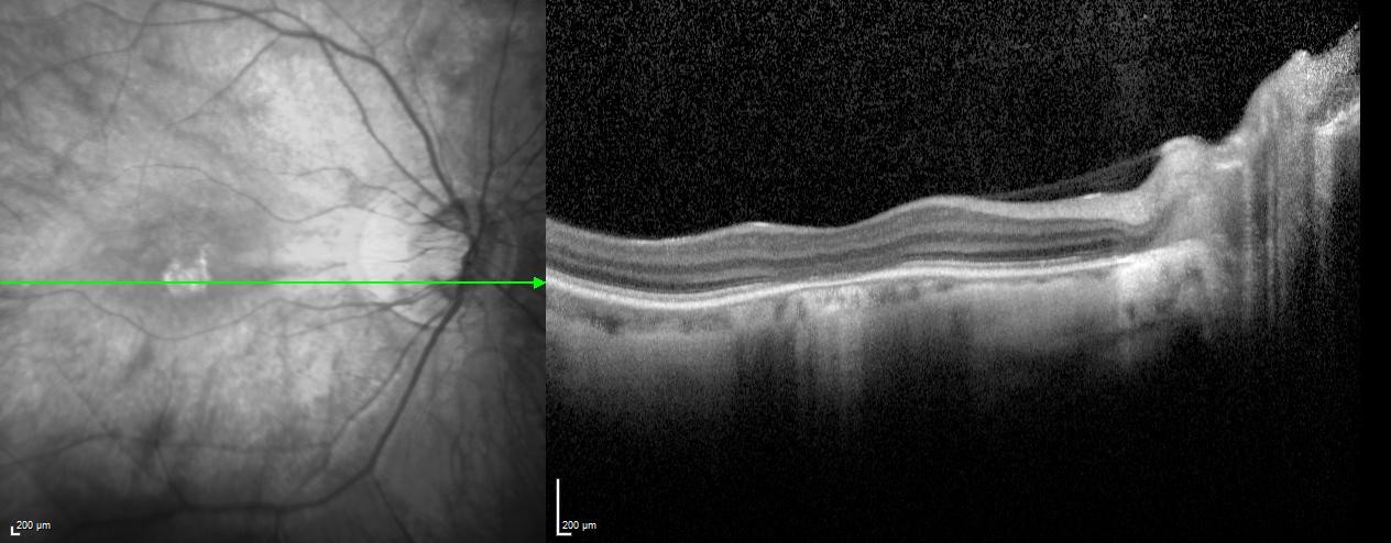
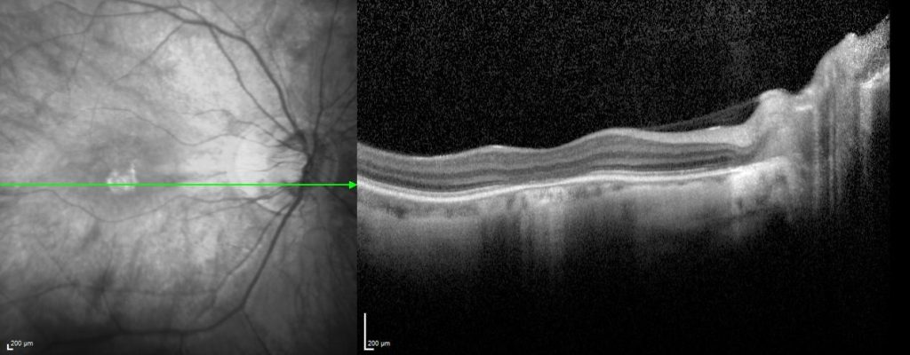
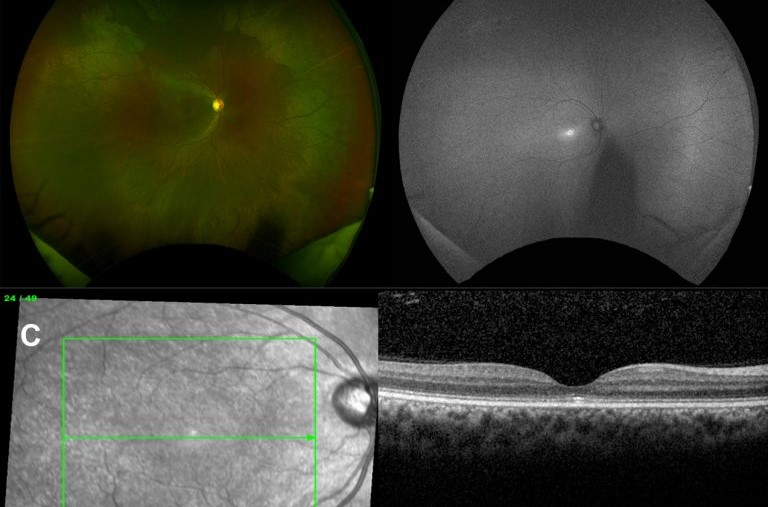
- Fundus Examination: Normal retinal appearance
Based on the clinical findings and history, the patient was diagnosed with Achromatopsia.

Achromatopsia Disease Entity
Achromatopsia is a rare, hereditary vision disorder characterized by a complete absence of color vision, reduced visual acuity, and increased sensitivity to light (photophobia).
This condition is also known as rod monochromatism or total color blindness. Patients with achromatopsia see the world in shades of gray due to the malfunctioning of cone cells in the retina.
Fundus Explorer Pro
Photograph the retinal findings described here with the phone already in your pocket — 22 D optics and built-in illumination in one handheld unit.
From Choroida — the team behind this sitePathophysiology
Achromatopsia is caused by genetic mutations that affect the cone cells in the retina. These cone cells are responsible for color vision and visual acuity.
There are three types of cone cells, each sensitive to different wavelengths of light (red, green, and blue). In individuals with achromatopsia, these cone cells are either absent or nonfunctional.
Instead, vision relies on the rod cells, which are more sensitive to light but do not detect color. This results in complete color blindness and other visual impairments.
Several genes have been identified in association with achromatopsia, including CNGA3, CNGB3, GNAT2, PDE6C, and PDE6H.
Mutations in these genes disrupt the normal functioning of cone cells, leading to the clinical manifestations of the disorder.
Achromatopsia Epidemiology
Achromatopsia is a rare condition, affecting approximately 1 in 30,000 individuals worldwide.
It is inherited in an autosomal recessive pattern, meaning both copies of the gene must be mutated for the condition to manifest.
It affects both genders equally and is typically diagnosed in early childhood when parents notice their child’s visual difficulties and unusual sensitivity to light.
Clinical Features
The clinical presentation of achromatopsia is typically evident from infancy or early childhood. Key features include:
- Color Blindness: Patients see the world in shades of gray, unable to distinguish any colors.
- Reduced Visual Acuity: Visual sharpness is significantly decreased, often ranging from 20/60 to 20/200.
- Photophobia: Extreme sensitivity to light, causing discomfort or pain in bright environments.
- Nystagmus: Involuntary, rapid eye movements that are common in individuals with achromatopsia.
- Normal Fundus Appearance: Despite significant visual impairment, the retina often appears normal on examination.

Achromatopsia Diagnosis
Diagnosing achromatopsia involves a combination of clinical examination, patient history, and specialized tests:
- Visual Acuity Testing: Assessing the sharpness of vision using standard eye charts.
- Color Vision Testing: Using tests such as the Ishihara plates to evaluate the ability to perceive colors.
- Electroretinography (ERG): Measuring the electrical responses of the retina to light stimuli. In achromatopsia, cone responses are absent or significantly reduced, while rod responses are typically normal.
- Genetic Testing: Identifying mutations in the associated genes can confirm the diagnosis and provide information for genetic counseling.
Differential Diagnosis
Several other conditions can present with similar symptoms, making differential diagnosis essential:
- Cone Dystrophy: A group of disorders characterized by progressive loss of cone cell function, leading to similar visual impairments.
- Leber Congenital Amaurosis (LCA): A severe retinal dystrophy that presents in infancy with poor vision and nystagmus.
- Stargardt Disease: An inherited macular dystrophy that causes progressive vision loss and difficulty in low-light conditions.
Achromatopsia Management
Currently, there is no cure for achromatopsia, but various management strategies can help improve the quality of life for affected individuals:
- Visual Aids: Low-vision aids such as magnifying glasses, telescopic lenses, and electronic devices can help with reading and other visual tasks.
- Tinted Lenses: Specially designed glasses with red or dark tints can reduce light sensitivity and improve comfort in bright environments.
- Adaptive Devices: Use of devices like screen readers and audiobooks to assist with daily activities.
- Gene Therapy: Experimental treatments aimed at restoring cone cell function are under investigation and hold promise for future therapeutic options.
Prognosis
The prognosis for individuals with achromatopsia is generally stable, with no progressive loss of vision over time.
However, the quality of life can be significantly affected due to visual impairments and light sensitivity.
Early diagnosis and appropriate management can help patients adapt to their condition and lead fulfilling lives.
Prevention and Public Health Implications
Since achromatopsia is a genetic disorder, there are no preventive measures. However, genetic counseling can help at-risk families understand the likelihood of passing the condition to their children.
Public health efforts should focus on raising awareness about the condition and promoting early diagnosis and intervention.
Conclusion
Achromatopsia is a rare, hereditary disorder that profoundly affects color vision and visual acuity. Understanding its pathophysiology, clinical features, and management options is crucial for improving patient outcomes.
While there is no cure, various strategies can help manage symptoms and improve quality of life. Advances in genetic research and potential gene therapies offer hope for future treatments.


Document what you see
Two smartphone imaging tools built for everyday clinic use — one for the slit lamp, one for the fundus.
From Choroida — the team behind this siteReferences
- Kohl, S., et al. (2005). Mutations in the CNGB3 gene encoding the beta-subunit of the cone photoreceptor cGMP-gated channel cause achromatopsia. American Journal of Human Genetics, 76(2), 380-389.
- Michaelides, M., et al. (2004). Achromatopsia: Progress in phenotype and molecular genetic studies. Journal of Medical Genetics, 41(7), 546-555.
- Sundin, O. H., et al. (2000). Genetic basis of total colourblindness among the Pingelapese islanders. Nature Genetics, 25(3), 289-293.
- Wissinger, B., et al. (2001). CNGA3 mutations in hereditary cone photoreceptor disorders. American Journal of Human Genetics, 69(4), 722-737.
- Zobor, D., et al. (2017). Gene therapy for cone photoreceptor disorders. Current Opinion in Ophthalmology, 28(4), 277-283.
