
CASE REPORT
A 28-year-old woman suffering from type 1 sialidosis, underwent a complete ophthalmological assessment at the eye clinic complaining of a progressive decline of visual acuity in recent months.
Polymerase chain reaction (PCR) and Sanger sequencing analysis of the NEU1 gene disclosed an already described compound heterozygosity. The patient suffered from myoclonic epilepsy with hypotonia and severe motor disability, from approximately the age of 13 years.
The best corrected visual acuity (BCVA) measured with the Early Treatment Diabetic Retinopathy Study (ETDRS) chart at 2 m was: 45 letters (20/63) in the right eye (OD) and 33 letters (20/100) in the left eye (OS).
Slit lamp examination showed a slight diffuse opacity of the corneal endothelium in both eyes, with incipient lens cortical opacities. There was no nystagmus.
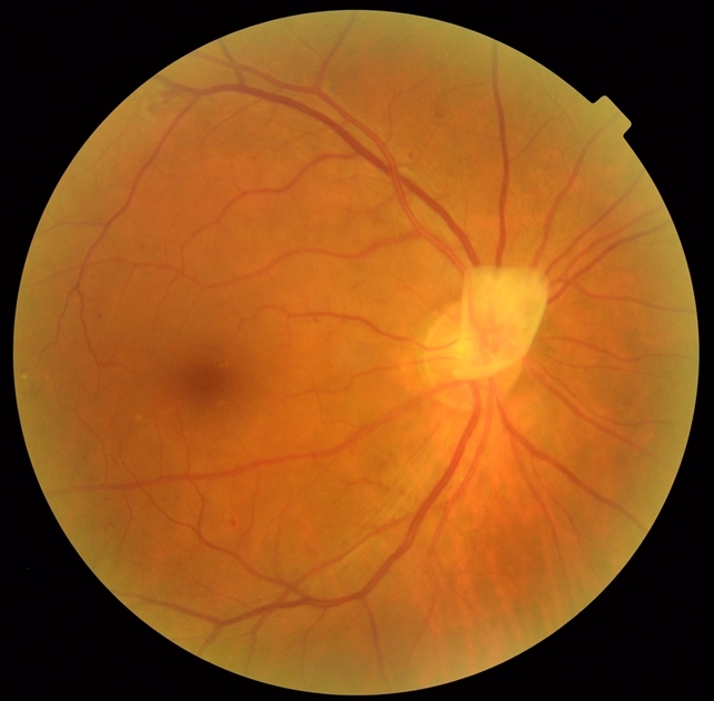
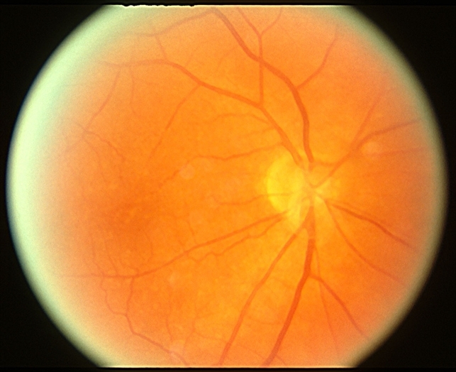
Fundus examination and retinography performed with True Color confocal ophthalmoscope EIDON, showed a macular cherry-red spot bilaterally, with diffuse dystrophy of the retinal pigment epithelium in the middle periphery.
In the right eye the optic disc was normal, while in the left eye, on the nasal portion of the optic disc, there was a whitish area of vitreous thickening, residual from the fetal vascularization on the optic disc ” Bergmeister’s papilla”.
DISEASE
Bergmeister’s papilla was first reported by Dr. Otto Bergmeister in 1877. Also referred to as an epipapillary veil, it is a remnant of the fibrous sheath surrounding the fetal hyaloid artery during embryological development.
In the fetus, blood is supplied to various parts of the eye by the hyaloid artery, which travels through the optic nerve head to the posterior aspect of the lens, where it branches into the tunica vasculosa lentis.
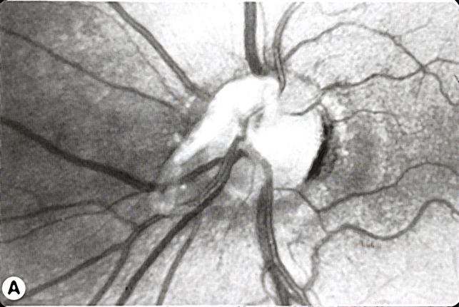
During normal development at around 30 weeks of gestational age, this blood supply is resorbed and develops into Cloquet’s (hyaloid) canal. Incomplete resorption of these structures during development can result in persistent fetal vasculature, such as the Mittendorf dot and the Bergmeister’s papilla.
It is suspected that defects in genes guiding cell apoptosis result in persistent fetal vasculature.
Fundus Explorer Pro
Photograph the retinal findings described here with the phone already in your pocket — 22 D optics and built-in illumination in one handheld unit.
From Choroida — the team behind this siteSymptoms
Bergmeister’s papilla is typically asymptomatic and found incidentally, changes to vision would depend upon association with secondary effects on the retina or macula.
Diagnostic procedures
Bergmeister’s papilla can be observed with ophthalmoscopy and fundus photography, but they are more likely to be identified through optical coherence tomography (OCT). Visualization reveals a cluster of veil-like glial or fibroglial tissue emerging anteriorly, centrally, and nasally from the optic nerve head.
The presence of papillae can influence the size of the optic nerve head cup. Bergmeister’s papilla can be unilateral or bilateral.

Papillae can be vascularized by a retinal vessel, otherwise known as a prepapillary vascular loop. If it is vascularized, this can be visualized on fluorescein angiography (FA). Without vascularization, the FA of a patient with Bergmeister’s papilla would be unremarkable.
MANAGEMENT
None indicated if Bergmeister’s papilla are without associated complications.


Document what you see
Two smartphone imaging tools built for everyday clinic use — one for the slit lamp, one for the fundus.
From Choroida — the team behind this siteREFERENCES
- Bergmeister O. Beiträge zur Entwicklungsgeschichte des Säugethierauges. Wien Embryologische Institut Mitteilungen. 1877;1:63-84.
- Royal Society of London. B. In: Catalogue of Scientific Papers (1800-1883) Supplementary Volume. Vol XII. Cambridge, UK: Cambridge University Press; 1902:72.
- Orellana J, Friedman AH. Bergmeister’s Papilla. In: Clinico-Pathological Atlas of Congenital Fundus Disorders. New York, New York: Springer; 1993:107-108. doi:10.1007/978-1-4613-9320-7_20
- Goldberg MF. Persistent fetal vasculature (PFV): An integrated interpretation of signs and symptoms associated with persistent hyperplastic primary vitreous (PHPV). Am J Ophthalmol. 1997;124(5):587-626. doi:10.1016/s0002-9394(14)70899-2
- Petersen HP. Persistence of the Bergmeister papilla with glial overgrowth. Acta Ophthalmol. 2009;46(3):430-440. doi:10.1111/j.1755-3768.1968.tb02826.x
- Lin Q, Deng J, Ohno-Matsui K, He X, Xu X. The existence and regression of persistent Bergmeister’s papilla in myopic children are associated with axial length. Transl Vis Sci Technol. 2021;10(13):4. doi:10.1167/tvst.10.13.4.
