Congenital toxoplasmosis is a leading infectious cause of retinochoroiditis in infants.
The parasite Toxoplasma gondii crosses the placenta after a primary maternal infection in pregnancy, and the retina is a frequent target, with the macula often involved. Therefore, every child born to an infected mother needs a dilated fundus exam.
How Congenital Toxoplasmosis Reaches the Eye
Maternal infection usually passes unnoticed, because most adults have mild or no symptoms. Then tachyzoites cross the placenta and spread to the fetal brain and retina. Transmission becomes more likely as pregnancy advances, although early fetal infection tends to cause more severe disease.
The Macular Scar in Congenital Toxoplasmosis
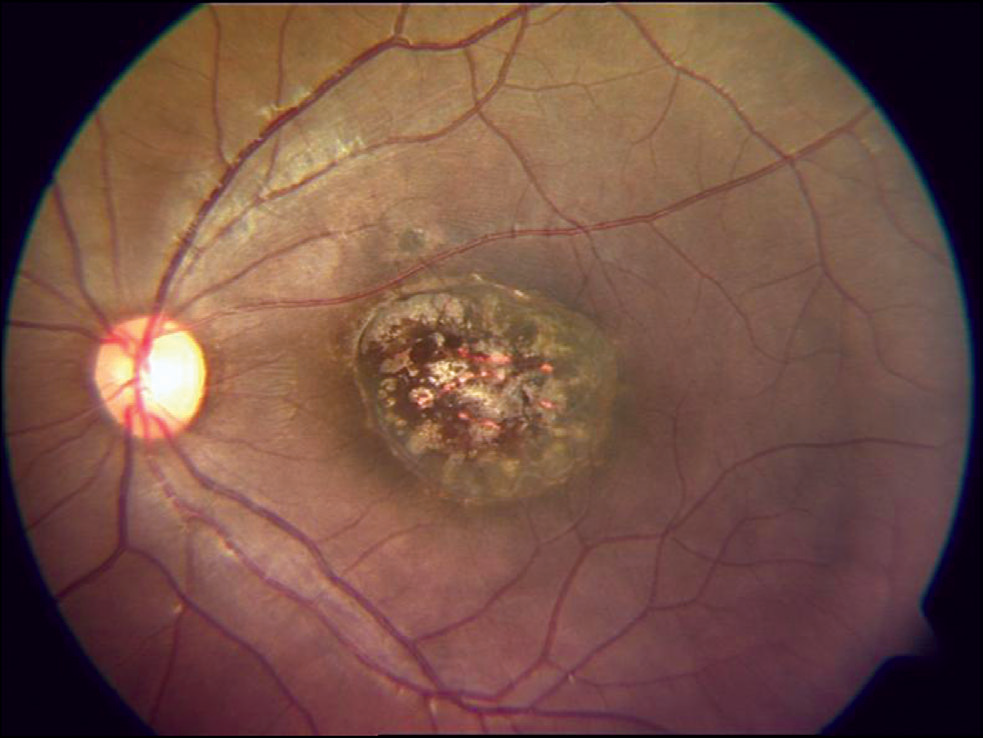
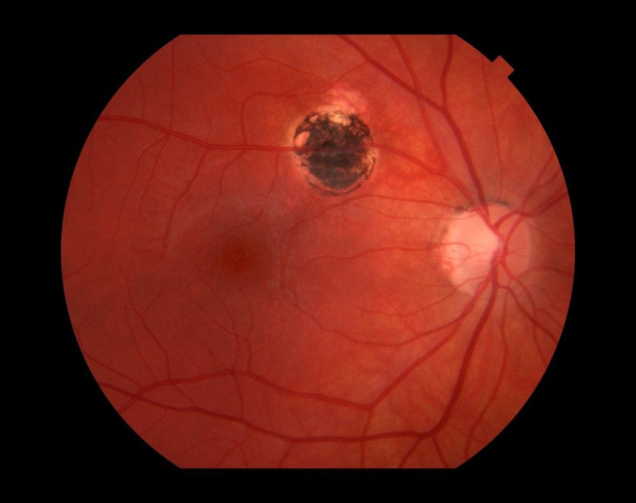
The typical lesion is a punched-out, atrophic retinochoroidal scar at the posterior pole, with a white center of bare sclera and dense pigment at the border.
Large scars can resemble a macular coloboma. Because the scar often involves the fovea, central vision in that eye is usually poor.
Fundus Explorer Pro
Photograph the retinal findings described here with the phone already in your pocket — 22 D optics and built-in illumination in one handheld unit.
From Choroida — the team behind this siteBilateral Disease in Congenital Toxoplasmosis
Congenital disease affects both eyes more often than acquired infection does, and one fundus may hold several scars, sometimes with small satellite lesions.
Bilateral macular scars carry the worst visual prognosis. Therefore, examine the second eye carefully even when one lesion explains the findings.
Associated Ocular Signs
- Strabismus, often the reason for the first referral
- Nystagmus with bilateral macular damage
- Microphthalmia after severe early infection
- Cataract, optic atrophy and anterior uveitis
Examining an Infant With Congenital Toxoplasmosis
First, dilate both pupils and use indirect ophthalmoscopy. Next, record the location, size and activity of each lesion. Wide-field photos help later comparison, and in addition a handheld slit lamp shows cells or cataract (see slit lamp technique).
Active Versus Healed Lesions
Active lesions look fluffy, white and poorly demarcated, and overlying vitritis may create the headlight in fog appearance.
Healed scars, by contrast, have sharp edges and pigmented margins. A fresh lesion beside an old scar suggests reactivation (see reading toxoplasmosis scars).
Imaging in Children
OCT of old scars shows retinal thinning, outer retinal loss and choroidal excavation, while active lesions show thickened, hyperreflective inner retina with vitreous cells above. However, infants rarely cooperate, so imaging may require examination under anesthesia.
Confirming Congenital Toxoplasmosis
Diagnosis combines serology with the clinical picture, and specific IgM or IgA in the infant supports congenital infection.
Also, IgG that persists beyond the first year confirms it, because maternal IgG fades by then. Before birth, amniotic fluid PCR can establish fetal infection.
Differential Diagnosis
- Congenital cytomegalovirus and other TORCH infections
- Toxocara granuloma
- Developmental macular coloboma
- Congenital hypertrophy of the retinal pigment epithelium
Treatment of the Infected Infant
Infants usually receive pyrimethamine, sulfadiazine and folinic acid, often for about one year. In a large US cohort, treated children appeared to develop fewer new lesions than historical untreated groups. However, drugs cannot restore vision lost to an established macular scar, and pyrimethamine suppresses the bone marrow, so pediatricians monitor blood counts regularly.
Prenatal Treatment and Its Limits
Clinicians may start spiramycin or pyrimethamine with sulfadiazine after maternal seroconversion. A meta-analysis suggested that earlier treatment may lower transmission, but evidence that it prevents eye lesions remains weak, and as a result practice varies between countries.
Late Reactivation in Congenital Toxoplasmosis
Dormant cysts persist in scar borders for life, and long-term cohort data show that new lesions, even a first lesion, can appear in adolescence. Still, most children in treated cohorts keep useful vision when one macula is spared.
Visual Prognosis in Congenital Toxoplasmosis
First, visual outcome depends mainly on whether the fovea is involved, so peripheral scars in both eyes often allow good acuity. However, a foveal scar leaves that eye with poor central vision, while a healthy fellow eye usually supports normal daily function. In addition, amblyopia therapy may help only when the scar spares the fovea.
Complications to Monitor
Next, check intraocular pressure, because uveitis can raise it. Also, choroidal neovascularization can develop at the edge of an old scar. Therefore, ask parents to report sudden squint or a white pupil without delay.
Follow-Up of Children Born to Infected Mothers
Examine the fundus at birth or soon after. Then repeat exams frequently in the first year and regularly through adolescence, and also check alignment and amblyopia at each visit.
Counseling Parents About Congenital Toxoplasmosis
Explain that old scars do not grow, while new lesions can appear later. Young children rarely complain of unilateral blur, so keep scheduled visits even when the child seems well, and finally teach older children to report floaters or blurred vision promptly.
Pitfalls to Avoid
- Do not assume a single scar means unilateral disease
- Never label a macular scar a coloboma without serology
- Do not stop follow-up after a normal newborn exam
- Avoid steroids without antiparasitic cover in active disease


Document what you see
Two smartphone imaging tools built for everyday clinic use — one for the slit lamp, one for the fundus.
From Choroida — the team behind this siteReferences
- Wallon M, Garweg JG, Abrahamowicz M, et al. Ophthalmic outcomes of congenital toxoplasmosis followed until adolescence. Pediatrics. 2014;133:e601-e608.
- McLeod R, Boyer K, Karrison T, et al. Outcome of treatment for congenital toxoplasmosis, 1981-2004: the National Collaborative Chicago-Based, Congenital Toxoplasmosis Study. Clin Infect Dis. 2006;42:1383-1394.
- SYROCOT Study Group, Thiébaut R, Leproust S, Chêne G, Gilbert R. Effectiveness of prenatal treatment for congenital toxoplasmosis: a meta-analysis of individual patients’ data. Lancet. 2007;369:115-122.
- Montoya JG, Liesenfeld O. Toxoplasmosis. Lancet. 2004;363:1965-1976.
Test yourself
A few questions straight from this article.
-
How does the timing of maternal infection affect congenital toxoplasmosis?
Tachyzoites cross the placenta after primary maternal infection; transmission becomes more likely as pregnancy advances, although early fetal infection tends to cause more severe disease. -
What is the typical fundus lesion of congenital toxoplasmosis?
The typical lesion is a punched-out, atrophic retinochoroidal scar at the posterior pole, with a white center of bare sclera and dense pigment at the border. -
A large congenital macular scar is found in an infant. Which developmental lesion can it resemble, requiring serology before labelling?
Large toxoplasma scars can resemble a macular coloboma, so a macular scar should never be labelled a coloboma without serology. -
How does an active toxoplasma retinochoroiditis lesion differ from a healed scar?
Active lesions look fluffy, white and poorly demarcated, with vitritis creating a headlight in fog appearance, while healed scars have sharp edges and pigmented margins. -
Which serological finding confirms congenital toxoplasmosis in an infant?
Specific IgM or IgA supports congenital infection, and IgG persisting beyond the first year confirms it because maternal IgG fades by then. -
Which regimen do infants with congenital toxoplasmosis usually receive, often for about one year?
Infants usually receive pyrimethamine, sulfadiazine and folinic acid for about a year; pyrimethamine suppresses the marrow, so blood counts are monitored. -
What is the strength of evidence that prenatal treatment after maternal seroconversion prevents eye lesions of congenital toxoplasmosis?
A meta-analysis suggested earlier treatment may lower transmission, but evidence that it prevents eye lesions remains weak, so practice varies. -
Why do children with congenital toxoplasmosis need follow-up through adolescence?
Dormant cysts persist in scar borders for life, and new lesions, even a first lesion, can appear in adolescence; old scars themselves do not grow. -
What mainly determines visual outcome in congenital toxoplasmosis?
Outcome depends mainly on foveal involvement: peripheral scars in both eyes often allow good acuity, while a foveal scar leaves that eye with poor central vision. -
Which vision-threatening complication can develop at the edge of an old congenital toxoplasma scar?
Choroidal neovascularization can develop at the edge of an old scar, and intraocular pressure should also be checked because uveitis can raise it.

Writes and reviews the clinical articles on Educate. Every post is checked against peer-reviewed sources before it goes live.