Hemangioma vs melanoma is a frequent referral question when an orange or nonpigmented choroidal mass appears near the macula.
Circumscribed choroidal hemangioma is a benign vascular hamartoma, but amelanotic melanoma can look strikingly similar. Therefore, the decision rests on color, ultrasound, angiography and growth.
Why Hemangioma vs Melanoma Causes Confusion
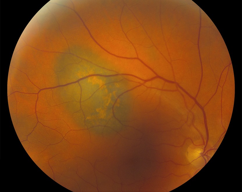
Both tumors arise in the choroid and can cause subretinal fluid, and many melanomas carry little pigment. Meanwhile, overlying RPE hyperplasia can darken part of a hemangioma, so color alone may mislead experienced examiners.
Patient Profile and Symptoms
Hemangioma usually presents in middle age with blurred vision, metamorphopsia or a hyperopic shift from macular fluid, while melanoma also affects adults, especially those with fair skin and light irides. However, small melanomas are often asymptomatic and found on routine examination, so the history rarely settles the diagnosis.
Fundus Explorer Pro
Photograph the retinal findings described here with the phone already in your pocket — 22 D optics and built-in illumination in one handheld unit.
From Choroida — the team behind this siteColor in Hemangioma vs Melanoma
A typical hemangioma is reddish orange, and its margins blend gradually into the normal fundus. By contrast, an amelanotic melanoma usually looks yellow, tan or creamy, often with mottled brown areas. Also, surface clumps of orange lipofuscin favor melanoma (see choroidal nevus vs small melanoma).
Shape and Location
Hemangiomas sit mostly in the posterior pole, often near the disc or fovea, and form a smooth, low dome a few millimeters thick. Melanoma can occur anywhere and may break through Bruch membrane, and that mushroom shape essentially excludes hemangioma.
Ultrasound in Hemangioma vs Melanoma
Ultrasound is the most useful single office test, and on B-scan and A-scan, a hemangioma shows uniform high internal reflectivity from its many vascular interfaces. Melanoma, however, shows low to medium internal reflectivity, acoustic hollowness, choroidal excavation and sometimes orbital shadowing, and then record thickness and basal diameter at every visit.
Fluorescein Angiography Findings
On FA, a hemangioma fills very early, often in the prearterial or arterial phase, with a lacy vascular pattern, and late frames then show diffuse staining. In contrast, a melanoma fills more slowly and patchily, and larger tumors may show a double circulation, but overlap is considerable, so FA rarely decides the case alone.
ICG Pattern in Hemangioma vs Melanoma
Indocyanine green angiography gives the most characteristic sign, because a hemangioma fills rapidly and intensely within the first minute. Then, in late frames, the dye washes out and the lesion turns relatively hypofluorescent. Melanoma usually fills slowly and variably, without this early bright, late washout sequence.
OCT and the Overlying Retina
Over a hemangioma, OCT often shows chronic intraretinal cysts and retinal edema, while melanoma more often shows fresh subretinal fluid with shaggy, elongated photoreceptors. Also, enhanced depth imaging may reveal large choroidal vessels inside a hemangioma, but these patterns overlap, so OCT supports the other tests.
Growth in Hemangioma vs Melanoma
Hemangiomas usually remain stable in size over long follow-up, and vision falls because of fluid while the tumor itself barely changes. Therefore, documented enlargement of a choroidal mass should raise strong concern for melanoma.
Other Mimics to Exclude
Several lesions also enter this differential:
- Choroidal metastasis, creamy yellow and often multifocal
- Choroidal osteoma, with bright plate-like echoes
- Posterior scleritis, with a T-sign on ultrasound
- Melanocytoma, dark and usually at the disc
Treatment in Hemangioma vs Melanoma
Symptomatic hemangioma is usually treated with verteporfin photodynamic therapy (see choroidal hemangioma). PDT dries the macula and often flattens the tumor, but it cannot reverse chronic photoreceptor loss.
Melanoma, however, needs plaque brachytherapy, proton beam therapy or enucleation (see choroidal melanoma). As a result, PDT for a misdiagnosed melanoma could delay effective treatment.
Counseling the Patient
Patients often hear the word tumor and assume cancer, so for hemangioma, explain that the lesion is benign and threatens vision only through fluid. Suspected melanoma, by contrast, calls for referral to an ocular oncologist and a discussion of systemic surveillance, and when doubt persists, say so and explain the monitoring plan.
Follow-Up Plan
First, document the lesion with photography, OCT and ultrasound at baseline, and next, recheck any uncertain lesion within a few months. Finally, growth, new orange pigment or new fluid should trigger urgent oncology referral.
Quick Table: Hemangioma vs Melanoma
- Color: reddish orange versus tan, yellow or mottled
- Ultrasound: high versus low to medium internal reflectivity
- Shape: low dome versus dome or mushroom
- ICG: early intense filling with late washout versus slow filling
- Growth: stable versus progressive
Pitfalls to Avoid
- Do not diagnose hemangioma on color alone
- Never start PDT for an orange mass without ultrasound
- Also, remember that RPE hyperplasia can darken a hemangioma
- Refer early when imaging findings conflict


Document what you see
Two smartphone imaging tools built for everyday clinic use — one for the slit lamp, one for the fundus.
From Choroida — the team behind this siteReferences
- Shields CL, Honavar SG, Shields JA, Cater J, Demirci H. Circumscribed choroidal hemangioma: clinical manifestations and factors predictive of visual outcome in 200 consecutive cases. Ophthalmology. 2001;108:2237-2248.
- Shields JA, Zimmerman LE. Lesions simulating malignant melanoma of the posterior uvea. Arch Ophthalmol. 1973;89:466-471.
- Shields CL, Shields JA, De Potter P. Patterns of indocyanine green videoangiography of choroidal tumours. Br J Ophthalmol. 1995;79:237-245.
- Shields CL, Manalac J, Das C, Ferguson K, Shields JA. Choroidal melanoma: clinical features, classification, and top 10 pseudomelanomas. Curr Opin Ophthalmol. 2014;25:177-185.
- Collaborative Ocular Melanoma Study Group. Accuracy of diagnosis of choroidal melanomas in the Collaborative Ocular Melanoma Study. COMS report no. 1. Arch Ophthalmol. 1990;108:1268-1273.
Test yourself
A few questions straight from this article.
-
What is a circumscribed choroidal hemangioma?
Circumscribed choroidal hemangioma is a benign vascular hamartoma, though amelanotic melanoma can look strikingly similar. -
What is the typical fundus colour of a circumscribed choroidal hemangioma?
A typical hemangioma is reddish orange and its margins blend gradually into the normal fundus, whereas amelanotic melanoma is yellow, tan or creamy. -
Which surface finding on an orange choroidal mass favours melanoma over hemangioma?
Surface clumps of orange lipofuscin favour melanoma; RPE hyperplasia, by contrast, can darken part of a hemangioma and mislead on colour. -
Which tumour shape essentially excludes a choroidal hemangioma?
Hemangiomas form a smooth, low posterior-pole dome; melanoma may break through Bruch membrane, and that mushroom shape essentially excludes hemangioma. -
What internal reflectivity does a choroidal hemangioma show on A-scan and B-scan ultrasound?
A hemangioma's many vascular interfaces give uniform high internal reflectivity, while melanoma shows low to medium reflectivity, hollowness and choroidal excavation. -
Which indocyanine green angiography sequence is most characteristic of choroidal hemangioma?
On ICG a hemangioma fills rapidly and intensely within the first minute, then washes out to relative hypofluorescence; melanoma fills slowly and variably. -
What does OCT of the overlying retina often show over a choroidal hemangioma?
Hemangiomas often show chronic intraretinal cysts and oedema, whereas melanoma more often shows fresh subretinal fluid with shaggy photoreceptors. -
Documented enlargement of a choroidal mass on follow-up should raise strong concern for which diagnosis?
Hemangiomas usually stay stable in size, with vision falling from fluid, so documented growth raises strong concern for melanoma. -
What is the usual treatment for a symptomatic circumscribed choroidal hemangioma?
Verteporfin PDT dries the macula and often flattens a hemangioma; melanoma instead needs brachytherapy, proton beam or enucleation, so misdiagnosis delays care. -
Which refractive change can macular fluid from a circumscribed choroidal hemangioma cause?
Hemangioma usually presents in middle age with blurred vision, metamorphopsia or a hyperopic shift from macular fluid.

Writes and reviews the clinical articles on Educate. Every post is checked against peer-reviewed sources before it goes live.