
Aggressive posterior ROP (AP-ROP) is a severe, rapidly progressive form of retinopathy of prematurity that develops in the posterior retina and does not move through the classic stages.
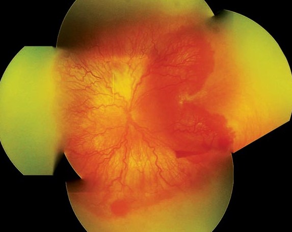
It was formerly called rush disease, and the name reflects the speed with which it can also progress to retinal detachment.
Because its signs differ from the usual ones, clinicians can miss it, and the consequences are serious.
What Is Aggressive Posterior ROP?
AP-ROP occurs in zone I or in the posterior part of zone II.
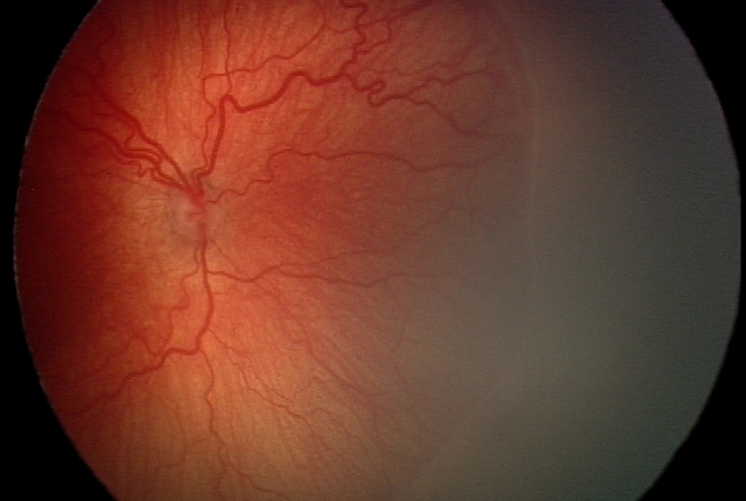
Also, the vessels in the posterior pole show severe dilation and tortuosity, out of proportion to the changes in the periphery.
The neovascularization is also flat, forming a network at the border of vascularized and avascular retina, and does not show the typical ridge of stage 2 or 3.
In addition, shunts and loops of vessels appear.
The disease can progress from early signs to retinal detachment within days to weeks.
The third edition of the International Classification of ROP retained the term and defined its features (see ROP screening).
Risk Factors of Aggressive Posterior ROP
- Extremely low gestational age and birth weight
- Prolonged oxygen exposure and fluctuations in oxygen levels
- Poor postnatal growth and severe systemic illness
- Occurrence in larger infants in resource-limited settings, where oxygen control is limited
It occurs in a small proportion of ROP, but it accounts for a high share of poor outcomes.
Fundus Explorer Pro
Photograph the retinal findings described here with the phone already in your pocket — 22 D optics and built-in illumination in one handheld unit.
From Choroida — the team behind this siteHow to Recognize It
- Plus disease that is present early and severe, with dilation and tortuosity in all four quadrants
- Poorly visible peripheral changes, since the retina is so immature
- A flat network of new vessels, not a ridge, which blends with the retina
- Vascular shunts and loops in the posterior pole
- Rapid change between examinations, so that the interval of one week is too long
The flat neovascularization is the feature that is most easily missed.
The examiner should look at the border between the vascular and avascular retina with care.
Wide-field imaging can also help to document the vessels and to follow changes.
Urgency
When you suspect AP-ROP, reexamine the infant within days.
Treatment should be planned without waiting for a classic stage.
The disease meets the criteria for type 1 ROP and treatment is indicated as soon as it is diagnosed (see retinopathy of prematurity).
Also, delay can be the difference between a treated eye and a detached retina.
Aggressive Posterior ROP: Treatment Options
- Laser photocoagulation: treats the avascular retina and reduces the stimulus for neovascularization
- Intravitreal anti-VEGF drugs: bevacizumab or ranibizumab, which reduce vessel proliferation rapidly and are particularly useful in zone I disease
- Combined treatment and vitrectomy: used for failures and for advanced stages (see ROP treatment)
The BEAT-ROP trial found that bevacizumab was more effective than laser for zone I stage 3 disease, with a lower rate of recurrence in zone I, though the study was not powered to show safety.
The RAINBOW trial compared ranibizumab with laser and found that treatment success was similar or better with ranibizumab.
Laser in zone I is technically hard, destroys much of the retina, and may produce a larger visual field loss, which is one reason for the interest in anti-VEGF therapy.
Concerns About Anti-VEGF Therapy
- Systemic absorption of the drug in a small infant and a possible effect on organ development
- Late recurrence of disease, requiring follow-up for months
- Uncertain long-term neurodevelopmental effects, which are under study
- Incomplete vascularization of the retina after treatment
For these reasons, the decision between laser and anti-VEGF depends on the zone, the infant’s condition, the availability of laser, and the team’s experience.
Inform parents of the risks and benefits, and obtain consent.
Follow-Up for Aggressive Posterior ROP
After treatment, examine the infant at frequent intervals for regression, recurrence, and complications.
Anti-VEGF therapy needs longer follow-up, often to well beyond the standard age, since reactivation can occur months later.
Refer older children for refraction, strabismus and amblyopia care, and long-term ophthalmic follow-up.
Differential Diagnosis
Other causes of retinal vessel abnormalities in infants include familial exudative vitreoretinopathy, Coats disease, and incontinentia pigmenti.
Consider them in unusual cases (see familial exudative vitreoretinopathy).
However, consider a premature baby with severe systemic illness and posterior vessel abnormalities to have AP-ROP until proven otherwise.
Working With the Neonatal Team
Communicate directly with the neonatologist, since the baby may be unstable, and the timing of treatment must fit the baby’s condition.
Clinicians may perform laser and injections at the bedside or in the operating room, with attention to anesthesia and monitoring.
Parents should be involved in the decisions, with a clear explanation of the urgency.
For the latest studies, see the PubMed results for aggressive posterior ROP.


Document what you see
Two smartphone imaging tools built for everyday clinic use — one for the slit lamp, one for the fundus.
From Choroida — the team behind this siteReferences
- Mintz-Hittner HA, Kennedy KA, Chuang AZ; BEAT-ROP Cooperative Group. Efficacy of intravitreal bevacizumab for stage 3+ retinopathy of prematurity. N Engl J Med. 2011;364:603-615.
- Stahl A, Lepore D, Fielder A, et al. Ranibizumab versus laser therapy for the treatment of very low birthweight infants with retinopathy of prematurity (RAINBOW): an open-label randomised controlled trial. Lancet. 2019;394:1551-1559.
- Chiang MF, Quinn GE, Fielder AR, et al. International Classification of Retinopathy of Prematurity, third edition. Ophthalmology. 2021;128:e51-e68.
- American Academy of Ophthalmology. Basic and Clinical Science Course, Section 6: Pediatric Ophthalmology and Strabismus. AAO; current edition.

Writes and reviews the clinical articles on Educate. Every post is checked against peer-reviewed sources before it goes live.


