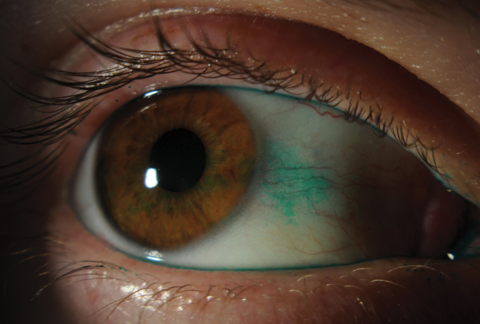
Lissamine green is a vital dye that stains conjunctival cells with damaged membranes or poor mucin cover.
Its blue-green signal stands out against the white sclera.
Therefore, clinicians pair it with fluorescein when they assess ocular surface disease.
However, the dye helps only when you apply it well and read it on time.
What Lissamine Green Actually Stains
Lissamine green is a synthetic organic acid dye.
It stains dead and degenerated epithelial cells.
It also marks cells that lack a healthy mucin glycocalyx.
In contrast, healthy epithelium stays unstained.
As a result, a stained area signals cellular barrier failure.
Why It Replaced Rose Bengal
Rose bengal produces a similar pattern but stings badly.
Lissamine green gives comparable staining with far better comfort.
As a result, most clinics now prefer it for conjunctival work.
All-fit Slit-Lamp Adapter
Record and share exactly what you see at the slit lamp. One adapter fits any slit lamp or surgical microscope — and any smartphone.
From Choroida — the team behind this siteLissamine Green Technique Step by Step
- First, finish noninvasive tests such as noninvasive tear breakup time.
- Next, wet a lissamine green strip with sterile saline.
- Then touch the strip to the inferior fornix.
- Finally, ask the patient to blink several times.
- Alternatively, instill a measured drop of 1% solution.
Dose Matters
A tiny drop on a strip may deliver too little dye.
Faint staining then looks normal.
Work on staining parameters suggests that dye volume affects the grade.
Therefore, keep the wetting volume consistent at every visit.
Timing the Lissamine Green Reading
Lissamine green staining fades within minutes.
Therefore, read the conjunctiva soon after instillation, usually within the first few minutes.
Reading immediately can also mislead, because excess dye pools in the tear film.
So wait for a few blinks, then grade promptly.
Light and Filters
Use white light at low to moderate intensity.
Bright light washes out faint spots.
Also, a red filter on the slit lamp can improve contrast.
Fluorescein Versus Lissamine Green
Fluorescein enters tissue where epithelial tight junctions break down.
It glows best on the cornea under cobalt blue light.
However, conjunctival fluorescein staining is hard to see without a yellow barrier filter.
Lissamine green, by contrast, gives sharp conjunctival contrast in white light.
Therefore, many clinicians grade the cornea with fluorescein and the conjunctiva with lissamine green (see corneal staining patterns).
Interpalpebral Pattern in Lissamine Green
Staining in the exposed nasal and temporal conjunctiva forms two triangles based at the limbus.
This pattern suggests aqueous deficient or evaporative dry eye.
In Sjögren syndrome, the staining can be dense and confluent.
Also, check the cornea for matching punctate fluorescein staining.
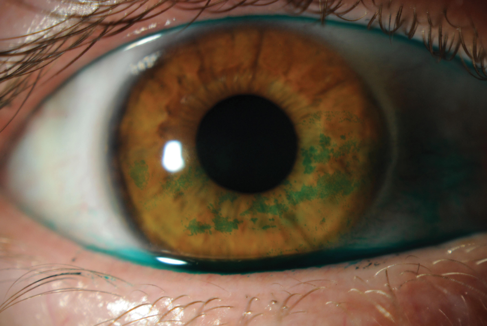
Superior Bulbar Staining
Staining above the limbus points away from simple dry eye.
First, lift the upper lid and look for superior limbic keratoconjunctivitis.
Next, consider contact lens irritation and floppy eyelid syndrome.
Also, ask about thyroid eye disease, which often accompanies SLK.
Inferior Staining With Lissamine Green
Inferior bulbar staining suggests exposure or incomplete closure.
Therefore, watch the blink and check for lagophthalmos.
Inferonasal staining may also reflect drop toxicity, because medications pool there.
In addition, conjunctivochalasis can stain along redundant folds.
Lid Margin and Lid Wiper Clues
Lissamine green outlines the line of Marx on the lid margin.
Anterior migration of this line can accompany meibomian gland dysfunction.
Next, evert the upper lid and inspect the lid wiper zone.
Staining there, called lid wiper epitheliopathy, correlates with dry eye symptoms in contact lens wearers.
Grading Lissamine Green Staining
Use one fixed scale at every visit.
The Oxford scheme compares cornea and conjunctiva against standard panels.
The van Bijsterveld score, first developed with rose bengal, grades nasal conjunctiva, temporal conjunctiva and cornea.
Also, the SICCA ocular staining score uses lissamine green for conjunctiva and fluorescein for cornea.
Lissamine Green in Dry Eye Diagnosis
TFOS DEWS II lists ocular surface staining as a homeostasis marker.
For example, more than nine conjunctival spots counts as positive.
Lid margin staining beyond set length and width thresholds also counts.
However, staining shows damage, while symptoms and tear tests complete the diagnosis.
When Staining Does Not Match Symptoms
Some patients report severe discomfort with minimal staining.
Then consider neuropathic pain, allergy or blepharitis.
Others show heavy staining with few symptoms.
As a result, test corneal sensation when staining far exceeds complaints.
Talking to the Patient
Warn that the eye may look green briefly.
Blinking clears the dye, and stinging is usually mild.
When possible, show the patient a photo of the stained areas.
Pitfalls to Avoid
- Reading too late, after the dye has faded
- Using a dry strip with a tiny drop
- Grading under bright light that hides faint spots
- Forgetting to lift the upper lid
- Calling inferior staining dry eye before checking closure


Document what you see
Two smartphone imaging tools built for everyday clinic use — one for the slit lamp, one for the fundus.
From Choroida — the team behind this siteReferences
- Manning FJ, Wehrly SR, Foulks GN. Patient tolerance and ocular surface staining characteristics of lissamine green versus rose bengal. Ophthalmology. 1995;102:1953-1957.
- Bron AJ, Evans VE, Smith JA. Grading of corneal and conjunctival staining in the context of other dry eye tests. Cornea. 2003;22:640-650.
- Whitcher JP, Shiboski CH, Shiboski SC, et al. A simplified quantitative method for assessing keratoconjunctivitis sicca from the Sjögren’s Syndrome International Registry. Am J Ophthalmol. 2010;149:405-415.
- Wolffsohn JS, Arita R, Chalmers R, et al. TFOS DEWS II diagnostic methodology report. Ocul Surf. 2017;15:539-574.
- Korb DR, Greiner JV, Herman JP, et al. Lid-wiper epitheliopathy and dry-eye symptoms in contact lens wearers. CLAO J. 2002;28:211-216.
Test yourself
A few questions straight from this article.
-
Which ocular surface cells does lissamine green stain?
Lissamine green stains dead and degenerated epithelial cells and cells that lack a healthy mucin glycocalyx, while healthy epithelium stays unstained, so staining signals barrier failure. -
Why has lissamine green largely replaced rose bengal for conjunctival staining?
Rose bengal produces a similar pattern but stings badly, whereas lissamine green gives comparable staining with far better comfort. -
When should lissamine green conjunctival staining be graded after instillation?
Lissamine green fades within minutes, but reading immediately misleads because excess dye pools in the tear film, so grade promptly after a few blinks. -
Which slit lamp setting improves the visibility of lissamine green staining?
Lissamine green is read in white light at low to moderate intensity because bright light washes out faint spots; a red filter can further improve contrast. -
When combining vital dyes on the ocular surface, how do many clinicians divide the grading between fluorescein and lissamine green?
Fluorescein glows best on the cornea under cobalt blue but conjunctival fluorescein is hard to see without a barrier filter, whereas lissamine green gives sharp conjunctival contrast in white light. -
Lissamine green forms two interpalpebral triangles based at the limbus. What does this pattern suggest?
Staining of the exposed nasal and temporal conjunctiva in two limbus-based triangles suggests aqueous deficient or evaporative dry eye. -
Lissamine green shows superior bulbar conjunctival staining above the limbus. What should be looked for first?
Superior staining points away from simple dry eye; lift the upper lid for superior limbic keratoconjunctivitis, then consider contact lens irritation and floppy eyelid syndrome. -
Inferior bulbar conjunctival staining with lissamine green most suggests which problem?
Inferior bulbar staining suggests exposure or incomplete closure, so watch the blink and check for lagophthalmos before calling it dry eye. -
In contact lens wearers, lissamine green staining of which upper lid zone correlates with dry eye symptoms?
After eversion of the upper lid, staining of the lid wiper zone, called lid wiper epitheliopathy, correlates with dry eye symptoms in contact lens wearers. -
Under TFOS DEWS II, which lissamine green conjunctival staining count is positive as a homeostasis marker?
DEWS II lists ocular surface staining as a homeostasis marker, with more than nine conjunctival spots counted as positive; symptoms and tear tests complete the diagnosis.

Writes and reviews the clinical articles on Educate. Every post is checked against peer-reviewed sources before it goes live.