
Slit-lamp exam is the most important piece of equipment in the present-day ophthalmologist’s armamentarium. It is a routine method to examine the eyes for any abnormalities or diseases.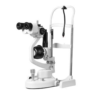

A slit lamp is composed of three basic parts:
- Observation system a low-power binocular microscope mounted horizontally.
- Illumination system consists of a bright soured of light, a condenser lens, a narrow variable slit, and a projecting lens.
- Mechanical system consists of a joystick and a vertically moveable chin rest for the patient. Joystick is used to move the microscope and illumination system towards or away from the eye and from side to side.
How to use Slit-lamp exam?
While performing Slit lamp exam following routine may be adopted:
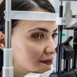
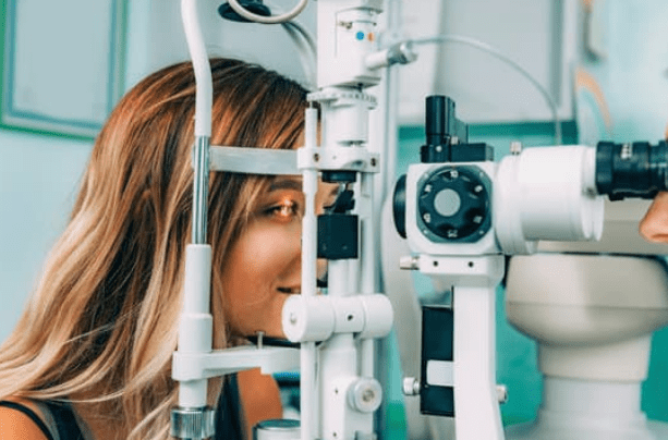
- The patient should be positioned comfortably in front of the slit lamp with his or her chin resting on the chin rest and forehead opposed to head support.
- The height of the table according to patient’s height.
- Examination should be carried out in semi dark room.
- Focus the microscope by
- Adjusting the interpupillary distance
- Adjusting the eyepieces (set at 0 or dial in your refraction)
- Set magnification in on 1x
- Adjust the illumination or brightness through the desired filters.
- Adjust the width and height of the beam.
The purpose of the Slit lamp examination is to permit the observer to examine the transparent media of the eye, that is, the cornea, the aqueous humor, the lens, and the vitreous.
The narrow beam provides an optical section of the transparent structures, which can be distinguished from one another by their differences in optical density.
The examiner usually views first the anterior segment of eye. The fundus examination by Slit lamp Exam is carried out by interposing an additional lens such as hruby lens (-55D), 90D or 78D lens.
The ophthalmologist will be able to detect many abnormal findings by carrying out the Slit-lamp exam such as
- Styes, folliculitis in eyelids
- Conjunctival congestion, presence of papilla and follicles in conjuctiva
- Corneal opacities & ulcers
- Neovascularization in iris in diabetic retinopathy or rubeosis iridis
- Abnormal reaction of pupil to light
- Cloudiness of lens (cataract)
- Vitreous floaters.
- Presence of foreign body
- Macular degeneration, retinal detachment and glaucomatous changes in optic disc and optic nerve.
- Damage to the retina and the blood vessels due to diabetes and hypertension.
Slit lamp examination uses
Modern Slit lamp smartphone photography with its auxiliary devices not only provides magnified views of every part of the eye from cornea to Retinal imaging.
but also Slit-lamp exam allows quantitative measurements (intraocular pressure, endothelial cell counts, pupil size, corneal thickness, anterior chamber depth, etc.)
Slit lamp photography of every part for documentation. Many ophthalmologists purchased slit-lamp cameras for photography which cost them too much.
The alternative is smartphone. smartphones Slit-lamp exam are readily available and are relatively inexpensive. It has a fully embedded system capable of acquisition, storage and analysis of fundus images that can be directly transmitted from the phone via the wireless telecommunication system.
The smartphones can be attached to slit-lamp with the help of slitlamp adapters.

