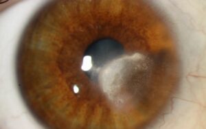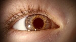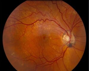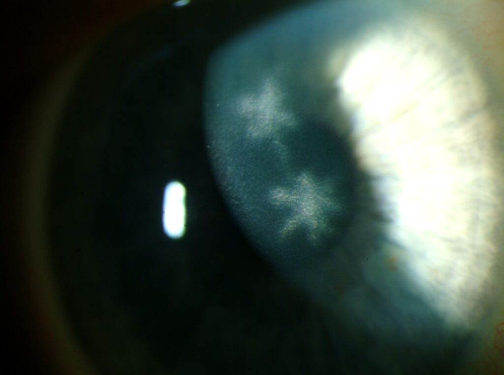
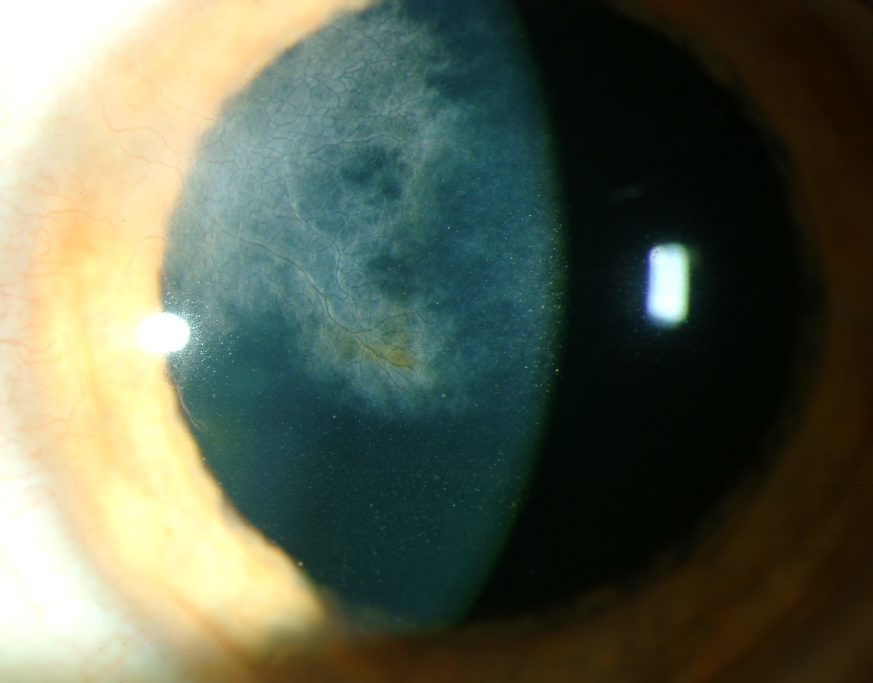
Corneal ghost vessels are empty stromal channels that remain after inflammatory vascularization subsides.
They appear as fine, pale, branching lines and carry little or no blood.
Because deep channels often follow interstitial keratitis, they can reveal an old, forgotten diagnosis.
What Corneal Ghost Vessels Look Like
Under direct illumination, they resemble thin gray threads within the stroma.
Retroillumination shows them well because the empty lumen stands out against the red reflex.
However, a narrow slit beam is needed to judge their depth.
Why Empty Vessels Persist
The healthy cornea suppresses vessel growth through antiangiogenic factors.
During inflammation, signals such as VEGF overcome that balance, and new vessels invade the stroma.
However, regression is often incomplete, so narrow channels can persist within the scarred stroma.
Mouse data also suggest that blood vessels regress less completely than lymphatics after brief inflammation.
All-fit Slit-Lamp Adapter
Record and share exactly what you see at the slit lamp. One adapter fits any slit lamp or surgical microscope — and any smartphone.
From Choroida — the team behind this siteCan Corneal Ghost Vessels Reperfuse?
Yes, recurrent inflammation can reopen existing channels, and blood may return quickly.
Therefore, regard these channels as dormant.
New injection with blood-filled vessels in an old scar suggests active disease.
Depth Points to the Cause
Superficial channels often follow pannus from contact lens wear, trachoma or chronic surface disease.
In contrast, deep channels just anterior to Descemet membrane suggest previous interstitial keratitis.
Sectoral mid-stromal channels beside a focal scar may follow herpetic disease.
Corneal Ghost Vessels and Interstitial Keratitis
Interstitial keratitis inflames the stroma and often spares the epithelium early.
Years later, the eye may look quiet, with only empty channels and patchy scars.
As a result, bilateral deep channels with diffuse scarring deserve a careful history and serology.
Causes to Consider
Congenital syphilis is the classic association, and it typically affects both eyes (see congenital syphilis keratitis).
Other causes include acquired syphilis, tuberculosis, herpes simplex, herpes zoster and Lyme disease.
Cogan syndrome also belongs on the list, especially with hearing loss or vertigo.
History and Companion Signs
First, ask about childhood eye inflammation, photophobia and maternal infection.
Hearing loss, notched incisors or a saddle nose suggest congenital syphilis.
Also test corneal sensation, because reduced sensation favors herpetic disease.
Imaging Corneal Ghost Vessels
Slit-lamp photography provides a useful baseline for later comparison.
Anterior segment OCT then shows the depth of associated scarring and thinning.
However, very slow flow may escape detection, so correlate any negative scan with the examination.
Testing After Finding Corneal Ghost Vessels
Unexplained bilateral deep channels justify syphilis testing.
Treponemal assays, such as FTA-ABS or TP-PA, usually stay reactive after past infection.
Meanwhile, nontreponemal tests such as RPR may be negative in late or treated disease.
Therefore, a negative RPR alone does not exclude old syphilis.
Add tuberculosis testing or audiometry when the history supports them.
Conditions That Mimic Empty Vessels
For example, lattice dystrophy produces refractile branching amyloid lines, often with recurrent erosions (see lattice dystrophy).
Prominent corneal nerves also branch, but they lack a limbal vascular origin.
Perfused vessels, by contrast, contain visible blood and may show leakage.
Effect on Vision
The channels themselves rarely limit vision.
Instead, reduced acuity usually comes from scarring, thinning or irregular astigmatism.
Therefore, corneal topography and a rigid contact lens trial can clarify the optical contribution.
Managing Corneal Ghost Vessels
Empty channels in a quiet eye need no direct treatment.
Topical steroids have no clear role without active inflammation, and they can raise intraocular pressure.
Anti-VEGF agents, fine-needle diathermy and laser target perfused vessels, although evidence remains limited.
Also, none of these options removes an established scar.
Confirmed syphilis requires systemic evaluation and treatment with an infectious disease team.
Corneal Ghost Vessels Before Keratoplasty
Stromal vascularization increases the risk of graft rejection (see graft rejection).
Therefore, map the depth and quadrants of all channels before keratoplasty.
However, surgical inflammation may reopen them, so monitor these grafts closely.
Follow-Up
Compare new photographs with the baseline at each visit.
Also look for injection, pain, stromal edema or fresh blood within old channels.
If you start steroids, check intraocular pressure regularly.
Counseling Patients With Corneal Ghost Vessels
Explain that these lines usually mark past inflammation and do not prove current infection.
A positive syphilis result requires a private discussion, because it can affect relatives and partners.
Finally, advise prompt review for a red, painful or newly blurred eye.
Pitfalls to Avoid
- Do not treat empty channels with steroids in a quiet eye
- Never rely on a negative RPR to exclude old syphilis
- Avoid blaming the channels for vision loss before assessing scar and astigmatism


Document what you see
Two smartphone imaging tools built for everyday clinic use — one for the slit lamp, one for the fundus.
From Choroida — the team behind this siteReferences
- Schwartz GS, Harrison AR, Holland EJ. Etiology of immune stromal (interstitial) keratitis. Cornea. 1998;17:278-281.
- Cursiefen C, Maruyama K, Jackson DG, et al. Time course of angiogenesis and lymphangiogenesis after brief corneal inflammation. Cornea. 2006;25:443-447.
- Azar DT. Corneal angiogenic privilege: angiogenic and antiangiogenic factors in corneal avascularity, vasculogenesis, and wound healing. Trans Am Ophthalmol Soc. 2006;104:264-302.
- Gupta D, Illingworth C. Treatments for corneal neovascularization: a review. Cornea. 2011;30:927-938.
- American Academy of Ophthalmology. Basic and Clinical Science Course, Section 8: External Disease and Cornea. San Francisco: AAO.

Writes and reviews the clinical articles on Educate. Every post is checked against peer-reviewed sources before it goes live.