Zoster keratitis is corneal disease caused by varicella zoster virus reactivation in the ophthalmic trigeminal division.
It can appear within days of the rash or emerge months later.
Because each corneal form has its own mechanism, treatment also differs.
Why Zoster Keratitis Takes Several Forms
Viral activity, immune inflammation and nerve damage can all injure the cornea.
Early epithelial lesions usually reflect viral activity, while stromal disease is largely immune mediated.
Later, denervation impairs surface healing.
Examining Zoster Keratitis
First, inspect the forehead, upper lid and tip of the nose.
Hutchinson sign raises the risk of ocular involvement because the nasociliary nerve also supplies the eye.
However, a clear nose does not exclude corneal disease.
Next, test corneal sensation before instilling anesthetic, then apply fluorescein.
Finally, measure intraocular pressure and look for anterior chamber cells.
All-fit Slit-Lamp Adapter
Record and share exactly what you see at the slit lamp. One adapter fits any slit lamp or surgical microscope — and any smartphone.
From Choroida — the team behind this sitePunctate Epithelial Lesions
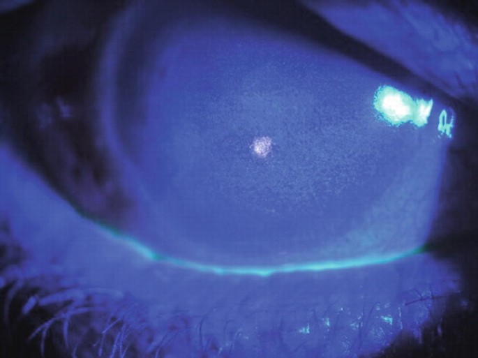
Coarse punctate lesions often appear first, within days of rash onset.
Patients usually report tearing, photophobia and foreign body sensation.
These foci may also coalesce into pseudodendrites.
Pseudodendrites in Zoster Keratitis
Pseudodendrites look like raised, ropy, gray plaques without terminal bulbs.
They also stain weakly with fluorescein.
In contrast, herpes simplex dendrites are true ulcers with terminal bulbs.
When the history is unclear, PCR from an epithelial sample can help separate the two viruses.
Delayed Mucous Plaques
Some patients develop branching plaques months after the acute illness.
These plaques adhere loosely to the epithelium and stain brightly with lissamine green.
Therefore, examine the whole cornea and avoid treating the plaque in isolation.
Nummular Anterior Stromal Keratitis
Fine granular infiltrates can form beneath Bowman layer, often about 10 days after the rash.
They likely reflect an immune response.
Topical steroids usually help when the infiltrates reduce vision.
In addition, the appearance overlaps with other causes of nummular keratitis, so history guides the diagnosis.
Disciform Zoster Keratitis
Disciform disease causes a disc of stromal edema, often with keratic precipitates and Descemet folds.
Because endothelial inflammation drives the edema, check the pressure at every visit.
Treatment usually combines topical steroids with oral antiviral cover.
Topical Steroids for Stromal Disease
Steroids reduce inflammation and may limit scarring.
However, they do not treat the virus or restore corneal nerves.
Their risks include raised pressure, cataract and delayed epithelial healing.
Therefore, confirm epithelial status first and taper slowly, because rebound inflammation is common.
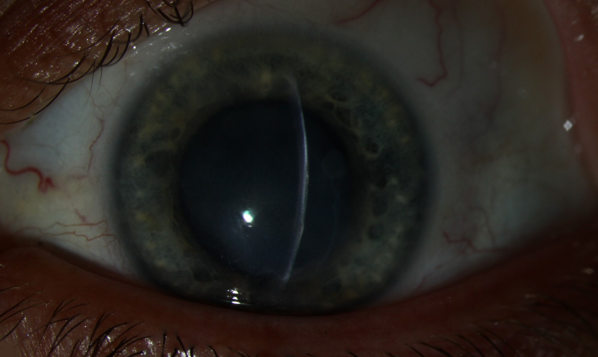
Antiviral Therapy for Zoster Keratitis
Oral therapy typically means valacyclovir 1 g or famciclovir 500 mg three times daily for 7 days.
Acyclovir 800 mg five times daily is an alternative, and renal function may require dose adjustment.
Treatment works best when started within 72 hours of rash onset.
Even so, antivirals cannot guarantee protection against later keratitis.
Long-Term Suppression in Zoster Keratitis
The Zoster Eye Disease Study tested one year of low-dose valacyclovir in selected patients with active ocular disease.
Clinicians should therefore review its published results before advising suppression.
Also, weigh age, renal function and recurrence history for each patient.
Neurotrophic Zoster Keratitis
Sensory loss reduces blinking, tear secretion and trophic support for the epithelium.
As a result, a large defect may cause surprisingly little pain.
Look for an oval epithelial defect with smooth, rolled edges.
Formal neurotrophic keratitis staging then helps guide treatment intensity.
Managing the Neurotrophic Cornea
First, stop unnecessary preserved drops and begin frequent preservative-free lubrication.
Next, consider autologous serum, a therapeutic contact lens, amniotic membrane or tarsorrhaphy for persistent defects.
Cenegermin, a recombinant nerve growth factor, improved healing in moderate and severe neurotrophic keratitis.
However, patients need frequent dosing, and access may be limited.
Lid Exposure and Corneal Thinning
Lid scarring, trichiasis and incomplete closure can worsen surface damage.
Progressive thinning in an anesthetic cornea can lead to melting and perforation.
Tissue adhesive, tarsorrhaphy or a patch graft may then preserve the globe.
Counseling Patients With Zoster Keratitis
Tell patients that chronic and recurrent disease can occur long after the rash heals.
They should report new redness, blur or discharge promptly, even without pain.
Also warn them against stopping steroids abruptly.
After recovery, discuss the shingles vaccine with the patient and primary care physician.
Pitfalls to Avoid
- Do not label every branching lesion as herpes simplex
- Never skip sensation testing before anesthetic drops
- Avoid steroids over an unexplained epithelial defect
- Do not rely on pain to judge severity


Document what you see
Two smartphone imaging tools built for everyday clinic use — one for the slit lamp, one for the fundus.
From Choroida — the team behind this siteReferences
- Liesegang TJ. Herpes zoster ophthalmicus natural history, risk factors, clinical presentation, and morbidity. Ophthalmology. 2008;115(2 Suppl):S3-S12.
- Cohen EJ, Kessler J. Persistent dilemmas in zoster eye disease. Br J Ophthalmol. 2016;100:56-61.
- Cohen EJ, Troxel AB, Liu M, et al. Zoster Eye Disease Study: rationale and design. Cornea. 2022;41:562-571.
- Bonini S, Lambiase A, Rama P, et al. Phase II randomized, double-masked, vehicle-controlled trial of recombinant human nerve growth factor for neurotrophic keratitis. Ophthalmology. 2018;125:1332-1343.
- American Academy of Ophthalmology. Basic and Clinical Science Course, Section 8: External Disease and Cornea. San Francisco: AAO.

Writes and reviews the clinical articles on Educate. Every post is checked against peer-reviewed sources before it goes live.