Retinal vessel caliber is the diameter of retinal arterioles and venules, and it reflects the state of the systemic microcirculation.
Population studies link narrow arterioles and wide venules to hypertension, stroke, and diabetes.
The measure has research value, but it is not yet a stand-alone test for individual risk.
How Retinal Vessel Caliber Is Measured
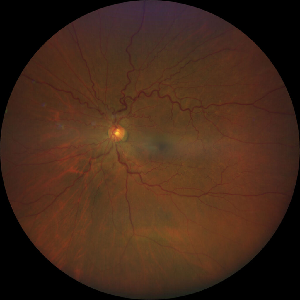
Software measures vessel widths on a disc-centered fundus photograph in a ring around the optic disc.
It summarizes them as the central retinal arteriolar equivalent and the central retinal venular equivalent.
Formulas by Parr and Hubbard, later revised by Knudtson, combine the measurements.
Image magnification, refraction, and the cardiac cycle can alter results, so studies use standard protocols.
Retinal Vessel Caliber and Blood Pressure
Wong and colleagues showed that narrower arterioles go with higher blood pressure and older age in a large population.
Sharrett and colleagues linked arteriolar narrowing to elevated pressure in the ARIC cohort.
Ikram and colleagues found that narrow arterioles predicted incident hypertension in the Rotterdam Study (see hypertensive retinopathy).
Fundus Explorer Pro
Photograph the retinal findings described here with the phone already in your pocket — 22 D optics and built-in illumination in one handheld unit.
From Choroida — the team behind this siteRetinal Vessel Caliber and Stroke
In the ARIC study, Wong and colleagues reported that retinal microvascular abnormalities predicted incident stroke.
Wider venules link to inflammation, obesity, and a higher risk of stroke in several cohorts.
These findings suggest that the retina mirrors cerebral small vessel disease.
Retinal Vessel Caliber in Diabetes and Kidney Disease
- Wider venules predict the development and progression of diabetic retinopathy
- Narrow arterioles associate with nephropathy
- Changes appear before overt retinal lesions in some studies
The relationships are statistical, so they apply to groups more than individuals (see diabetic retinopathy).
Retinal Vessel Caliber in Clinical Practice
Doctors rarely measure caliber by hand, since software is needed for reliability.
Be alert to generalized arteriolar narrowing in patients with uncontrolled blood pressure.
Photographs from screening programs may someday support cardiovascular risk estimates through automated methods.
Limits
Variation between devices, software, and imaging conditions limits clinical use.
Smoking, refractive error, and retinal disease alter the values.
Therefore, treat the result as supporting information.
An Example Case
A 62-year-old man with long-standing hypertension has a routine fundus photograph.
Software measures a central retinal arteriolar equivalent in the lowest quartile, and the venular equivalent is high.
The pattern fits with chronic blood pressure strain and raises concern about small vessel disease.
His physician reviews his blood pressure control and adds stroke prevention measures.
Questions Patients Ask
Patients ask whether the eye can show their heart risk, and the answer is that retinal vessels reflect small vessel health.
They also ask whether the eye test replaces a blood test, and it does not.
Because the data come from groups, explain that individual predictions are limited.
Pitfalls to Avoid
- Using a single measurement to predict risk in a patient
- Comparing values across studies with different software
- Forgetting the effect of refractive error and magnification
- Overinterpreting small differences between eyes
Looking Ahead
Deep learning models read fundus photographs for cardiovascular risk and may outperform simple caliber measures.
Also, large biobanks provide data to test these tools.
Therefore, retinal photographs may support population risk screening.
Practical Points
Capture disc-centered photographs of good quality.
Then use validated software with a documented protocol.
Also pair findings with blood pressure, glucose, and lipid data.
What Narrow Arterioles Mean
Narrow arterioles suggest long-standing hypertension and small vessel remodeling.
They also relate to future stroke and coronary events in cohort studies.
Because the change develops slowly, early control of blood pressure matters.
What Wide Venules Mean
Wide venules link to inflammation, hypoxia, obesity, and diabetes.
They predict stroke and diabetic retinopathy progression in some cohorts.
Therefore, venular widening may be a marker of metabolic stress.
Measuring Caliber at the Slit Lamp
Clinicians sometimes judge arteriolar narrowing by eye with the arteriole-to-venule ratio.
However, this estimate is subjective and varies between examiners.
Therefore, use photographs and software when you need a precise number.
Because small changes matter, avoid relying on a visual impression.
Linking the Eye to Systemic Care
Share notable findings with the patient’s physician, especially when blood pressure is poorly controlled.
Then record the finding and the plan in the chart.
Also reinforce that the eye examination supports, but does not replace, systemic screening.
Summary
Caliber is a research biomarker of systemic vessel health.
Therefore, rely on blood pressure and laboratory tests for decisions.
Also use the retinal finding as a prompt to review risk factors with the patient.
For the latest studies, see the PubMed results for retinal vessel caliber.


Document what you see
Two smartphone imaging tools built for everyday clinic use — one for the slit lamp, one for the fundus.
From Choroida — the team behind this siteReferences
- Wong TY, Klein R, Klein BEK, et al. Retinal vessel diameters and their associations with age and blood pressure. Invest Ophthalmol Vis Sci. 2003;44:4644-4650.
- Wong TY, Klein R, Couper DJ, et al. Retinal microvascular abnormalities and incident stroke: the Atherosclerosis Risk in Communities Study. Lancet. 2001;358:1134-1140.
- Ikram MK, Witteman JC, Vingerling JR, et al. Retinal vessel diameters and risk of hypertension: the Rotterdam Study. Hypertension. 2006;47:189-194.
- American Academy of Ophthalmology. Basic and Clinical Science Course, Section 12: Retina and Vitreous. AAO; current edition.

Writes and reviews the clinical articles on Educate. Every post is checked against peer-reviewed sources before it goes live.