Glaucoma suspect is the label for an eye with findings that raise concern for glaucoma, such as a suspicious optic disc, a raised pressure, or an equivocal field, without proof of glaucomatous damage.
It is also a common situation in clinic.
The task is to decide who needs treatment now, who needs close observation, and who can also be reassured (see primary open-angle glaucoma).
Who Becomes a Glaucoma Suspect
Suspicion arises from one or more findings:
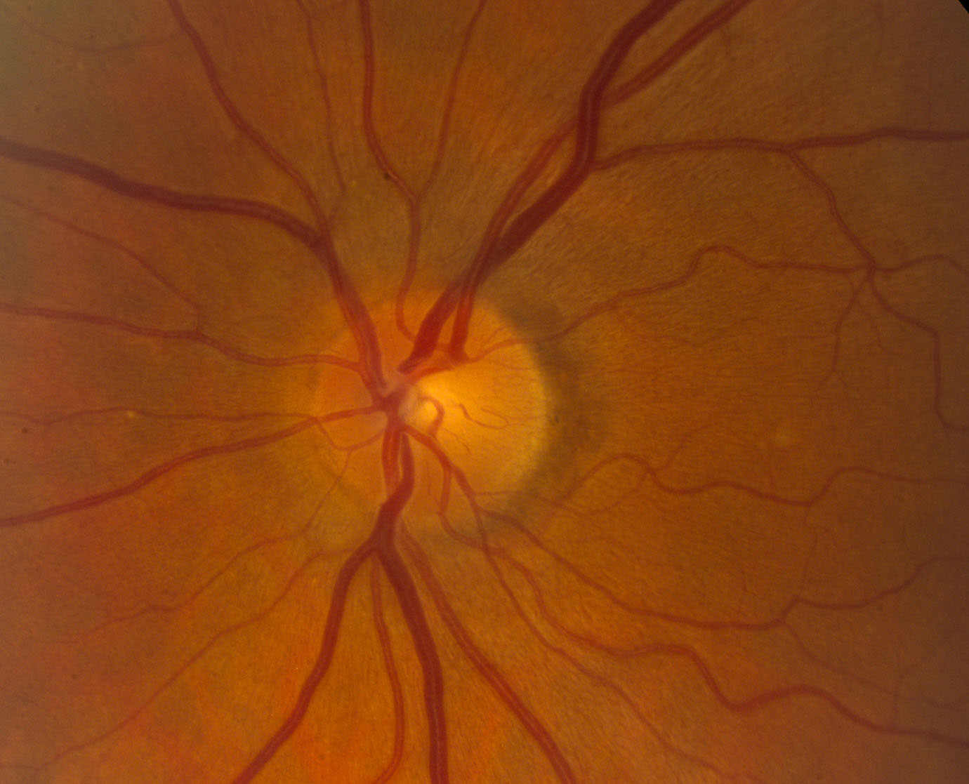
- A vertical cup-to-disc ratio above about 0.6, or an asymmetry of 0.2 or more between the eyes
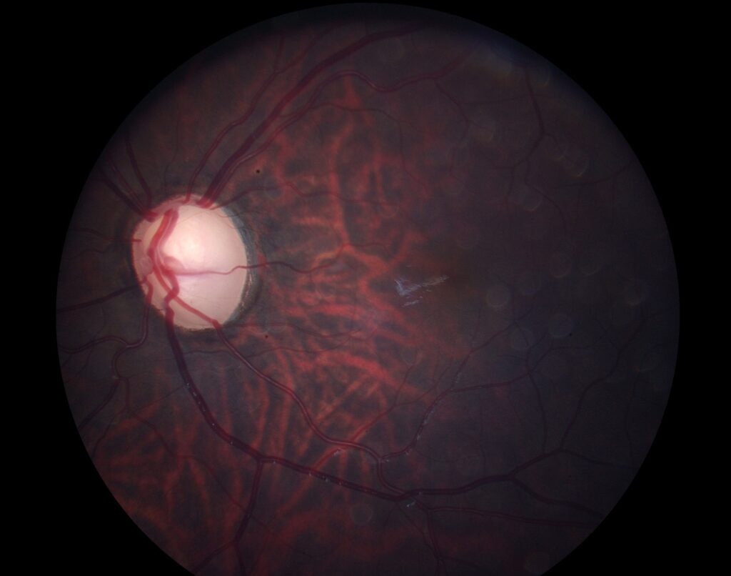
- A focal notch, a thin rim, or a disc hemorrhage (see optic disc hemorrhage in glaucoma)
- Retinal nerve fiber layer defect on examination or OCT
- Intraocular pressure above 21 mmHg on repeated measures (see ocular hypertension)
- A suspicious but not diagnostic visual field
Many large discs look cupped and are normal, so one finding alone is not enough.
Glaucoma Suspect: Risk Factors for Conversion
The Ocular Hypertension Treatment Study found that older age, higher pressure, thinner corneas, a larger vertical cup-to-disc ratio.
A higher pattern standard deviation predicted conversion (see the references).
Also, other risk factors include a family history, African ancestry, myopia, pseudoexfoliation, and low perfusion pressure.
A thin cornea reads low on applanation.
A thick one reads high, so pachymetry belongs in the first visit (see corneal pachymetry).
All-fit Slit-Lamp Adapter
Record and share exactly what you see at the slit lamp. One adapter fits any slit lamp or surgical microscope — and any smartphone.
From Choroida — the team behind this siteBaseline Examination
- Slit lamp examination and applanation tonometry (see Goldmann applanation tonometry)
- Gonioscopy to exclude angle closure (see gonioscopy)
- Dilated disc examination with stereo photographs
- Central corneal thickness
- Automated visual field, repeated if abnormal (see Humphrey visual field interpretation)
- OCT of the retinal nerve fiber layer and the macular ganglion cell complex
These tests form the baseline, because change is judged against them.
Glaucoma Suspect: Imaging and Function Together
Structure often changes before the field does, so OCT and disc photographs may show early damage when the field is normal.
A normal OCT does not exclude disease in a very large or very small disc.
Findings that agree across tests are also more reliable than one abnormal result.
Deciding Whether to Treat
Treat when the estimated risk of conversion is high, or when the patient is likely to have a long life with the disease.
The OHTS risk calculator combines age, pressure, thickness, cup-to-disc ratio, and pattern standard deviation to estimate the five-year risk.
A risk above about 15 percent is often used as a reason to consider treatment.
Clinicians can observe low-risk patients, and the decision should consider the patient’s age, health, and wishes.
Glaucoma Suspect: Follow-Up Schedule
A common plan is a review every six to twelve months, with the field and OCT yearly at first, and more often when the risk is high or the disc is changing.
Repeat the baseline tests after a few months to confirm stability, since learning effects and variability make the first results unreliable.
A new disc hemorrhage, a change in the rim, or a pressure rise should prompt a review of the plan.
Counselling
Explain that a suspect has no damage yet, that most suspects never develop glaucoma, and that follow-up is the safeguard.
Give a clear schedule and the reasons for it, and ask the patient to tell relatives about the risk of glaucoma in the family.
An Example Case
A 52-year-old man of African ancestry has a pressure of 25 mmHg in both eyes, a central corneal thickness of 520 micrometers.
A vertical cup-to-disc ratio of 0.65 in the right eye and 0.5 in the left.
The angle is open, the field is normal, and OCT shows a borderline thin inferior rim in the right eye.
His OHTS risk estimate is high, so he starts a prostaglandin analog, gets repeat baseline tests in three months, and returns every six months.
Mistakes to Avoid
- Calling a large physiologic cup glaucoma and starting treatment without other findings
- Failing to repeat an abnormal field before acting
- Missing narrow angles in a patient who is labeled as an open-angle suspect
- Stopping follow-up after one normal year, since conversion can occur late
What to Tell the Patient
Explain that being a suspect is not a diagnosis, that the eye is being watched for the earliest changes, and that treatment can start once the risk is clear.
Give a written schedule of visits and tests, and make sure that the patient understands why a missed visit matters.
Encourage relatives older than 40 to have an eye examination, since a family history raises the risk.
Newer Tools
Artificial intelligence reading of disc photographs and OCT may help to flag suspects, and home tonometry and wearable perimetry may give more data between visits.
Researchers are still testing these tools, and the standard of care remains a careful clinical evaluation with repeated measurements.


Document what you see
Two smartphone imaging tools built for everyday clinic use — one for the slit lamp, one for the fundus.
From Choroida — the team behind this siteReferences
- Gordon MO, Beiser JA, Brandt JD, et al. The Ocular Hypertension Treatment Study: baseline factors that predict the onset of primary open-angle glaucoma. Arch Ophthalmol. 2002;120:714-720.
- Prum BE Jr, Rosenberg LF, Gedde SJ, et al. Primary open-angle glaucoma preferred practice pattern guidelines. Ophthalmology. 2016;123:P41-P111.
- Weinreb RN, Aung T, Medeiros FA. The pathophysiology and treatment of glaucoma: a review. JAMA. 2014;311:1901-1911.
- American Academy of Ophthalmology. Basic and Clinical Science Course, Section 10: Glaucoma. AAO; current edition.

Writes and reviews the clinical articles on Educate. Every post is checked against peer-reviewed sources before it goes live.