Orbital apex syndrome is optic neuropathy combined with ophthalmoplegia from a lesion at the orbital apex.
The apex is a tight cone where the optic canal and superior orbital fissure meet, so a small lesion there can damage vision and eye movement together. Because several causes threaten sight or life, it needs a same-day workup.
Anatomy Behind Orbital Apex Syndrome
The optic nerve and ophthalmic artery pass through the optic canal. Next to it, the superior orbital fissure carries cranial nerves III, IV and VI, along with the ophthalmic division of the trigeminal nerve and sympathetic fibers. As a result, one lesion can produce a predictable cluster of deficits.
Clinical Signs of Orbital Apex Syndrome
- Reduced visual acuity and color vision
- Relative afferent pupillary defect
- Ophthalmoplegia, often complete
- Ptosis from third nerve involvement
- Forehead and corneal hypoesthesia from V1
- Mild proptosis in some cases
All-fit Slit-Lamp Adapter
Record and share exactly what you see at the slit lamp. One adapter fits any slit lamp or surgical microscope — and any smartphone.
From Choroida — the team behind this siteWhat the Patient Reports
Most patients describe painful, blurred or lost vision in one eye. They also notice double vision, although ptosis may hide it, and then numbness over the forehead often appears. However, pain varies, and some tumors present with a painless decline.
Fundus Findings in Orbital Apex Syndrome
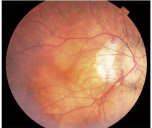
The optic disc often looks normal at first because the damage is posterior, and pallor then develops over several weeks. Disc edema suggests a more anterior or congestive process, while you should also check for retinal artery occlusion, which mucormycosis can cause.
Orbital Apex Syndrome vs Superior Orbital Fissure Syndrome
Superior orbital fissure syndrome shares the motor and V1 deficits, but vision and afferent pupil function stay normal. Therefore, the optic nerve marks the difference, and once an afferent defect appears, the lesion has reached the apex.
Comparison With Cavernous Sinus Syndrome
Cavernous sinus lesions also cause multiple ocular motor palsies, and they may add V2 sensory loss and Horner syndrome, while vision is usually spared early. Bilateral signs also favor the sinus, because it connects across the midline (see cavernous sinus cranial nerve patterns).
Urgent Causes of Orbital Apex Syndrome
Infection comes first in a diabetic or immunosuppressed patient, and mucormycosis and invasive aspergillosis spread from the sinuses into the apex. Bacterial orbital cellulitis can also extend posteriorly, and you should consider fractures and retrobulbar hemorrhage when the history fits.
Mucormycosis: The Emergency Not to Miss
Suspect it with diabetic ketoacidosis, recent corticosteroids or neutropenia, and look for black eschar on the palate or nasal turbinates. However, eschar is a late sign, so early nasal endoscopy helps. Delay costs vision and life, so involve ENT the same day (see mucormycosis case and management).
Other Causes
Inflammatory causes include IgG4-related disease, sarcoidosis and granulomatosis with polyangiitis, and Tolosa-Hunt syndrome and idiopathic orbital inflammation also occur. Neoplasms include lymphoma, metastases, meningioma and perineural spread of skin cancer, and finally, apex fractures can coexist with traumatic optic neuropathy.
Imaging in Orbital Apex Syndrome
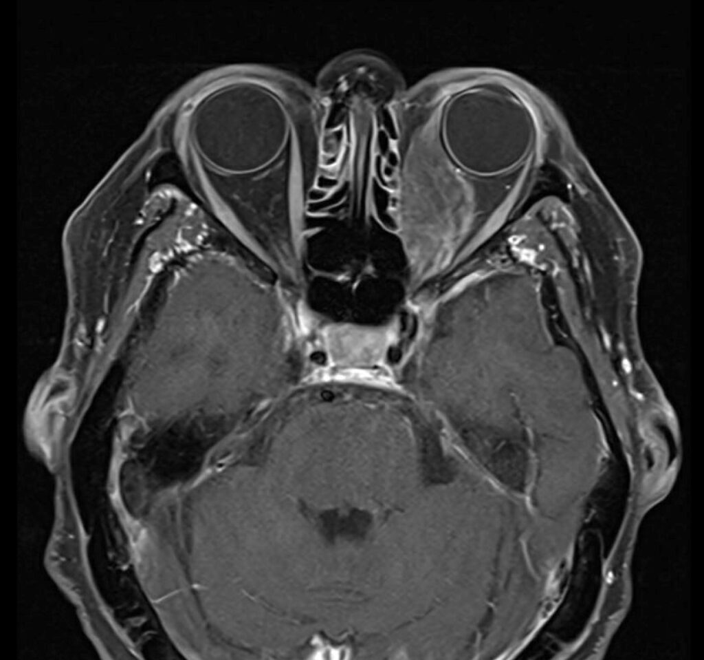
Order contrast MRI of the orbits and cavernous sinus with fat suppression, while CT shows bone erosion, fractures and sinus disease better. Diffusion-weighted imaging can also reveal infarction or necrotic fungal tissue, but a normal scan does not exclude early fungal infection.
Laboratory Workup
Check blood glucose, ketones and a complete blood count, then add ESR, CRP, ANCA and serum IgG4 when you suspect inflammation. In patients over 50, giant cell arteritis also deserves consideration.
Why Biopsy Matters in Orbital Apex Syndrome
Imaging rarely gives a final diagnosis, so tissue confirms fungus, lymphoma or granulomatous disease. Endoscopic biopsy through the sinus often reaches the apex safely. Because steroids can mask lymphoma and worsen fungal infection, biopsy first when feasible.
Treatment Principles
Treatment targets the cause, since no single therapy fits every patient. For mucormycosis, give liposomal amphotericin B and debride necrotic tissue urgently, and correct ketoacidosis and reduce immunosuppression where possible. Teams may discuss exenteration when the orbit is necrotic, although indications vary between centers, while inflammatory disease often responds to corticosteroids, then steroid-sparing agents.
Steroids in Orbital Apex Syndrome
A quick steroid response may tempt a diagnosis of Tolosa-Hunt syndrome, but lymphoma and sarcoidosis can also improve briefly. Therefore, treat it as a diagnosis of exclusion and repeat imaging, and withhold empirical steroids until you reasonably exclude infection.
Counseling and Prognosis
Explain that vision may not recover even after you control the cause, although motor palsies may improve more than optic nerve function. Warn about relapse in inflammatory disease, and next, arrange close follow-up with acuity, color vision, fields and motility.
Pitfalls to Avoid
- Do not label painful ophthalmoplegia as a diabetic palsy when vision is reduced
- Never give steroids before excluding fungal infection
- Do not rely on a normal disc in acute optic neuropathy
- Do not miss an afferent pupillary defect in a ptotic eye
- Never accept a normal scan if signs progress


Document what you see
Two smartphone imaging tools built for everyday clinic use — one for the slit lamp, one for the fundus.
From Choroida — the team behind this siteReferences
- Yeh S, Foroozan R. Orbital apex syndrome. Curr Opin Ophthalmol. 2004;15:490-498.
- Badakere A, Patil-Chhablani P. Orbital apex syndrome: a review. Eye Brain. 2019;11:63-72.
- Goyal P, Lee S, Gupta N, et al. Orbital apex disorders: imaging findings and management. Neuroradiol J. 2018;31:104-125.
- Cornely OA, Alastruey-Izquierdo A, Arenz D, et al. Global guideline for the diagnosis and management of mucormycosis. Lancet Infect Dis. 2019;19:e405-e421.
Test yourself
A few questions straight from this article.
-
Which structures pass through the optic canal at the orbital apex?
The optic nerve and ophthalmic artery pass through the optic canal, while the adjacent superior orbital fissure carries nerves III, IV and VI, V1 and sympathetic fibres. -
What distinguishes orbital apex syndrome from superior orbital fissure syndrome?
Superior orbital fissure syndrome shares the motor and V1 deficits but spares vision and afferent pupil function; once an afferent defect appears the lesion has reached the apex. -
Which features favour a cavernous sinus lesion over orbital apex syndrome?
Cavernous sinus lesions may add V2 sensory loss and Horner syndrome, usually spare vision early, and can be bilateral because the sinus connects across the midline. -
How does the optic disc usually look early in orbital apex syndrome?
Because the damage is posterior, the disc often looks normal at first and pallor develops over several weeks; disc edema suggests a more anterior or congestive process. -
Why is early nasal endoscopy advised when mucormycosis is suspected in orbital apex syndrome?
Mucormycosis is suspected with ketoacidosis, recent steroids or neutropenia; black eschar is a late sign, so early nasal endoscopy and same-day ENT input help. -
Which imaging is ordered first to assess the soft tissues in orbital apex syndrome?
Contrast MRI of the orbits and cavernous sinus with fat suppression is ordered, while CT shows bone erosion, fractures and sinus disease better. -
Why is biopsy preferred before steroids in orbital apex syndrome when feasible?
Imaging rarely gives a final diagnosis, and because steroids can mask lymphoma and worsen fungal infection, tissue is obtained first when feasible. -
Which drug is given for mucormycosis causing orbital apex syndrome?
Mucormycosis is treated with liposomal amphotericin B and urgent debridement of necrotic tissue, with correction of ketoacidosis and reduced immunosuppression. -
Why is Tolosa-Hunt syndrome treated as a diagnosis of exclusion in orbital apex syndrome?
A quick steroid response may tempt a Tolosa-Hunt diagnosis, but lymphoma and sarcoidosis can also improve briefly, so imaging is repeated and infection excluded first. -
Which deficit tends to recover least after the cause of orbital apex syndrome is controlled?
Vision may not recover even after the cause is controlled, and motor palsies may improve more than optic nerve function.

Writes and reviews the clinical articles on Educate. Every post is checked against peer-reviewed sources before it goes live.