
CASE REPORT
A 40 years old Chinese man sought treatment for the progressive deterioration of vision in the right eye over about five months period. His sister also had congenital ocular abnormalities.

On examination,
the Snellen visual acuity of the right eye and left eye was 0.15 and 0.6, respectively. The intraocular pressure of the right eye and the left eye were 11.3 and 12.7 mmHg, respectively.
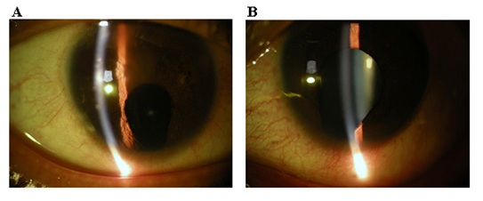
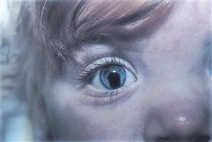
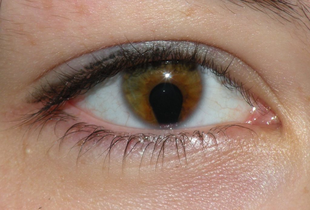
Slitlamp examination showed clear corneas in both eyes. Obviously, the patient had a bilateral inferonasal coloboma of the iris, which resulted in a pear-shaped pupil in both eyes.
DISEASE
Coloboma is derived from the Greek koloboma, meaning mutilated, curtailed, or with a defect. The term is used to describe ocular defects of the eyelids, iris, lens, ciliary body, zonules, choroid, retina, or optic nerve.
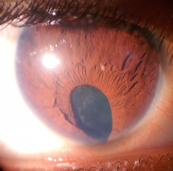
It is typically located in the inferonasal quadrant of the involved structure and is often associated with microphthalmia. It can affect one eye (unilateral) or both eyes (bilateral).
It is important to differentiate colobomas involving the globe from those of the eyelids. In either case, they can affect one eye (unilateral) or both eyes (bilateral).
MANAGEMENT
The most important predictor of visual outcome is the identification of normal foveal anatomy.
Patients with bilateral uveal coloboma or unilateral coloboma plus one other systemic abnormality should be referred to a genetics specialist to evaluate for systemic disorders.
Monocular precautions should be strongly considered for any patient with unilateral coloboma in an ophthalmic exam and resulting in decreased visual acuity on the affected side.
Interval monitoring for retinal detachment should be done with a dilated fundus exam approximately every 6-12 months or sooner if indicated for patients with posterior coloboma.
The risk of retinal detachment is there which may be up to 40%. Prophylactic laser of such eyes may reduce the occurrence of retinal detachment, though a randomized trial for this is not yet available.
Measures such as patching should be taken to maximize the visual potential of the affected side as there is often a normal retina present and refractive error is often present putting patients at risk for amblyopia in the ophthalmic exam.
HOW TO TAKE SLIT-LAMP EXAM IMAGES BY A SMARTPHONE?
Smartphone slit-lamp photography is the new advancement in the field of science and technology in which the photographs of the desired slit-lamp finding can be taken with smartphones by using the slit-lamp adapters.

REFERENCES
- Optic cup and stalk with open embryonic fissure below.
- Hyatt, G. A. & Dowling, J. E. Retinoic acid. A key molecule for eye and photoreceptor development. Invest. Ophthalmol. Vis. Sci. 38, 1471–1475 (1997).
- Nadauld, L. D. et al. Dual roles for adenomatous polyposis coli in regulating retinoic acid biosynthesis and Wnt during ocular development. Proc. Natl. Acad. Sci. U. S. A. 103, 13409–13414 (2006).
- Hornby, S. J., Ward, S. J. & Gilbert, C. E. Eye birth defects in humans may be caused by a recessively-inherited genetic predisposition to the effects of maternal vitamin A deficiency during pregnancy. Med. Sci. Monit. Int. Med.J. Exp. Clin. Res. 9, HY23–26 (2003).
