

Case Study
A 24-year-old woman presented for a routine eye examination. She reported no visual complaints and had no significant medical or family history.

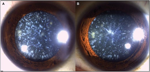
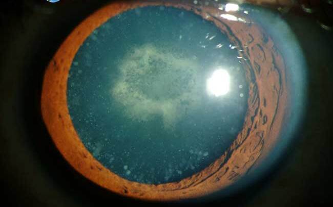
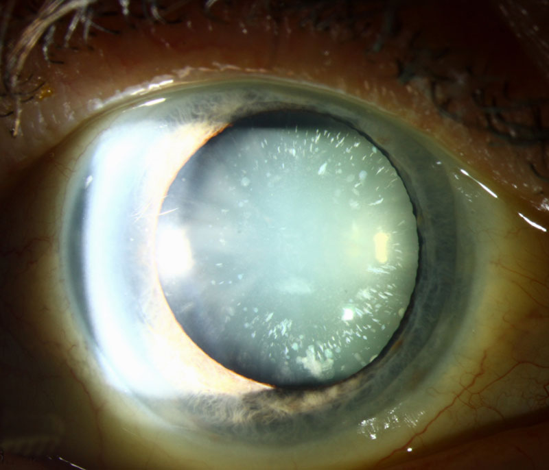
On slit-lamp examination, bilateral, small, bluish-white opacities were observed in the superficial layers of her lens cortex.
Her visual acuity was 20/20 in both eyes, and there was no evidence of glare or night vision difficulties. The findings were consistent with a diagnosis of cerulean cataract, a rare congenital lens anomaly.
Genetic testing revealed a mutation in the CRYBB2 gene, confirming its hereditary nature. Given her asymptomatic status, no intervention was necessary at the time, and regular follow-up was recommended.
Disease Entity
Cerulean cataract, also known as congenital blue-dot cataract, is a rare hereditary lens opacity characterized by distinct bluish-white or blue-gray opacities scattered in the cortex of the lens.
These opacities are often small and rounded, resembling dots or snowflakes, and are distributed symmetrically in both eyes.
Cerulean cataracts are typically congenital and non-progressive but may occasionally develop or worsen in adulthood.
While many individuals remain asymptomatic, severe cases can cause visual disturbances and require intervention.
Pathophysiology
The formation of cerulean cataracts is linked to genetic mutations affecting the crystalline proteins, which are essential for maintaining the transparency and refractive properties of the lens.
Specifically, mutations in the CRYBB2 and CRYGD genes, which encode beta- and gamma-crystallins, disrupt the structural organization of lens proteins, leading to the aggregation of opacities in the lens cortex.
These mutations are most commonly inherited in an autosomal dominant pattern, although autosomal recessive inheritance has been reported in some cases.
Environmental factors and oxidative stress may also contribute to the pathogenesis of cerulean cataracts.
Epidemiology
- Prevalence: Cerulean cataracts are rare, with no precise data on their prevalence. They are considered a subtype of congenital cataracts, which occur in approximately 1–6 per 10,000 live births.
- Inheritance: Most cases of cerulean cataracts are inherited in an autosomal dominant manner, although sporadic cases without a clear family history have been reported.
- Demographics: The condition affects males and females equally and is present at birth or develops early in life.
Clinical Features
The clinical presentation of cerulean cataracts can vary depending on the severity of the opacities and their impact on visual function.
- Characteristic Opacities: Bluish-white or gray dots scattered in the lens cortex are the hallmark feature. These opacities may appear clustered or distributed uniformly throughout the lens.
- Symmetry: The opacities are typically bilateral and symmetric, affecting both eyes equally.
- Vision: Most patients have normal or near-normal visual acuity, especially in mild cases. However, significant opacities may cause glare, blurred vision, or reduced contrast sensitivity.
- Stability: The cataracts are usually non-progressive or progress slowly over time.
Diagnosis
The diagnosis of cerulean cataracts is primarily clinical and based on characteristic findings during slit-lamp examination.
- Slit-Lamp Examination: Reveals distinct bluish-white opacities in the lens cortex. These opacities are most prominent in the superficial cortical layers and can be easily distinguished from other types of congenital cataracts.
- Genetic Testing: Testing for mutations in the CRYBB2 and CRYGD genes can confirm the diagnosis in hereditary cases.
- Family History: A thorough family history can help identify autosomal dominant inheritance patterns.
- Functional Assessment: Visual acuity, contrast sensitivity, and glare testing may be conducted to evaluate the impact of the cataracts on visual function.
Differential Diagnosis
Cerulean cataracts must be differentiated from other types of congenital or acquired cataracts with similar appearances.
- Coronary Cataracts: These involve spoke-like opacities near the lens periphery but lack the bluish hue of cerulean cataracts.
- Lamellar Cataracts: Present as concentric opacities in the lens nucleus or cortex, typically associated with metabolic conditions or systemic disorders.
- Blue-Dot Nuclear Cataracts: Rarely, other types of congenital cataracts may exhibit blue-dot opacities, but they are typically confined to the nucleus rather than the cortex.
- Cataracts Associated with Systemic Diseases: Conditions like Down syndrome, galactosemia, or Wilson’s disease can cause cataracts with similar appearances but are accompanied by systemic signs.

Management
Management of cerulean cataracts depends on the severity of the condition and its impact on visual function.
- Observation: In asymptomatic or mild cases, regular follow-up is recommended to monitor for progression.
- Glasses or Contact Lenses: Refractive correction may be sufficient for patients with mild visual disturbances.
- Surgical Intervention: Cataract surgery is indicated in cases with significant visual impairment affecting daily activities. Modern techniques like phacoemulsification with intraocular lens implantation can restore visual function effectively.
- Genetic Counseling: Families of affected individuals may benefit from genetic counseling to understand inheritance patterns and the risk of transmission to offspring.
Prognosis
The prognosis for individuals with cerulean cataracts is generally excellent, particularly in cases where the condition is mild and non-progressive.
- Visual Stability: Most individuals retain stable vision throughout their lives without the need for intervention.
- Surgical Outcomes: For those requiring cataract surgery, outcomes are typically favorable, with high rates of visual restoration.
- Quality of Life: Early diagnosis and appropriate management ensure that patients can maintain a good quality of life despite the condition.
Conclusion
Cerulean cataracts are a rare form of congenital lens opacity characterized by bluish-white cortical opacities. While most cases are asymptomatic and non-progressive, severe cases may require surgical intervention to restore vision.
Advances in genetic testing have improved our understanding of the condition’s pathogenesis and inheritance, allowing for better diagnosis and family counseling.
With proper management, individuals with cerulean cataracts can achieve excellent visual outcomes and lead normal lives.
HOW TO TAKE SLIT-LAMP EXAM IMAGES WITH A SMARTPHONE?
Smartphone slit-lamp photography is the new advancement in the field of science and technology in which photographs of the desired slit-lamp finding can be taken with smartphones by using the slit-lamp adapters.
Slit-lamp Smartphone photography
References
- Rahi JS, Sripathi S, Gilbert CE, Foster A. Childhood cataract: Epidemiology, management, and outcomes. Indian Journal of Ophthalmology. 1999;47(2):51-61.
- Berry V, Francis PJ, Kaushal S, et al. Missense mutations in CRYBB2 and CRYGD are associated with congenital cerulean cataracts. Human Molecular Genetics. 2001;10(9):949-955.
- Hejtmancik JF. Congenital cataracts and their molecular genetics. Seminars in Cell & Developmental Biology. 2008;19(2):134-149.
- Shiels A, Hejtmancik JF. Genetic origins of cataract: Insight from mouse models. Nature Reviews Genetics. 2007;8(7):522-532.
- Kausar T, Bhatti N, Ali M. Congenital cataract: Recent developments. Journal of Pediatric Ophthalmology & Strabismus. 2011;48(4):221-229.
