Rods vs cones is the basic contrast between the two photoreceptor types of the human retina.
Rods serve vision in dim light, while cones serve daylight acuity and color. This difference explains why some diseases start with night blindness and others start with central blur.
Structure: Rods vs Cones
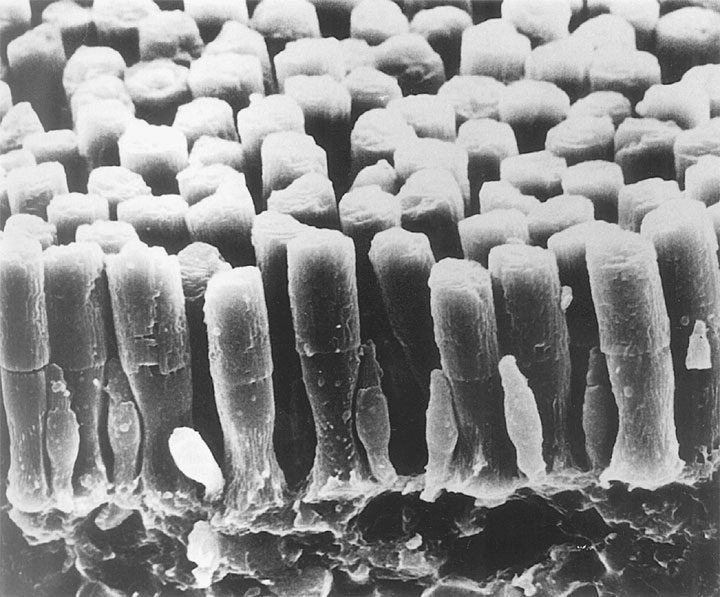
Both cells have an outer segment, an inner segment, a nucleus and a synaptic terminal. Rods have a long cylindrical outer segment filled with stacked, separate discs, while cones have a shorter, tapering outer segment, and their membrane folds stay open to the extracellular space.
Photopigments in Rods vs Cones
Rods contain rhodopsin, which absorbs best near 500 nm, while cones carry one of three opsins, each tuned to short, medium or long wavelengths. As a result, color vision needs at least two cone classes working together, and a single rod class cannot signal hue, so night vision stays colorless.
Fundus Explorer Pro
Photograph the retinal findings described here with the phone already in your pocket — 22 D optics and built-in illumination in one handheld unit.
From Choroida — the team behind this siteNumbers and Distribution
Curcio and colleagues counted about 92 million rods and 4.6 million cones in an average human retina. Cone density peaks at the foveal center, which contains no rods, while rod density, by contrast, peaks in a ring outside the macula. Therefore, a faint star is easier to see when you look slightly beside it.
Rods vs Cones in the Fovea
The foveal pit holds tightly packed, slender cones, and each central cone links to its own midget bipolar and ganglion cell pathway. This private wiring gives high spatial resolution, and consequently, visual acuity falls sharply even a few degrees from fixation.
Sensitivity and Convergence
Many rods converge onto a single ganglion cell, and because the cells pool their signals, rods can detect single photons. However, pooling sacrifices spatial detail, and rods also saturate in ordinary daylight, so cones carry vision in bright conditions.
Dark Adaptation: Rods vs Cones
Cones recover sensitivity quickly after a bright light, then reach a plateau, and next, after several minutes, rods take over at the rod-cone break. Full rod sensitivity then takes about 30 to 40 minutes, and a delayed rod phase suggests rod dysfunction or a visual cycle defect.
The Visual Cycle
After light bleaches rhodopsin, the retinal pigment epithelium regenerates the chromophore, and rods depend heavily on this RPE pathway, which includes RPE65. Evidence suggests cones also draw on a Müller cell pathway, which may explain their faster recovery, and therefore, visual cycle defects often present as slow dark adaptation.
Symptoms of Rod-First Disease
Patients report poor night vision and trouble moving in dim rooms, and then they notice they bump into objects at their sides. Central acuity often stays useful until late, and retinitis pigmentosa is the classic example.
Rod-First Disease on Fundus Examination
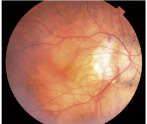
Typical RP shows bone spicule pigment in the midperiphery, arteriolar attenuation and waxy disc pallor, while cystoid macular edema and posterior subcapsular cataract may also reduce vision. On OCT, the ellipsoid zone shrinks from the periphery toward the fovea, although early disease can show little pigment at all.
Symptoms of Cone-First Disease
Cone and cone-rod dystrophies begin with reduced central vision and photophobia, and color discrimination also deteriorates early. Patients often see better in dim light, but later, rod loss adds night blindness and peripheral field loss (see cone-rod dystrophy).
Cone-First Disease on Examination
The macula may look normal at first, but then bull’s eye maculopathy or macular atrophy can develop. OCT shows foveal ellipsoid zone disruption, while the periphery stays intact for longer, and achromatopsia, a congenital cone disorder, adds nystagmus and severe light sensitivity.
Rods vs Cones on the Full-Field ERG
The ISCEV standard separates rod and cone function under controlled conditions, and the dark-adapted 0.01 response isolates rods. Light-adapted single flash and 30 Hz flicker responses reflect cone pathways, while the dark-adapted strong flash combines both systems (see electroretinography).
ERG Patterns in Rods vs Cones Disease
In RP, rod responses fall more than cone responses, and implicit times are often delayed. Cone dystrophy reverses this pattern, with reduced cone responses and relatively preserved rods, and in achromatopsia, cone responses are absent while rod responses stay normal.
Stationary vs Progressive Disorders
Night blindness does not always mean degeneration, for example in many forms of congenital stationary night blindness, which show a negative ERG (see congenital stationary night blindness). In addition, history and serial testing separate stable from progressive disease.
Counseling on Rods vs Cones Disease
Rods work at night and in the periphery, while cones handle faces and print, so explain which system matches the patient’s symptoms. Also discuss night driving, home lighting, tinted lenses for photophobia, and low vision services.
Pitfalls to Avoid
- Do not call every case of night blindness retinitis pigmentosa
- Also check for vitamin A deficiency after bariatric surgery or with malabsorption
- Do not rely on a normal fundus to exclude cone dysfunction
- Record the ERG under ISCEV conditions with adequate dark adaptation
- Remember that macular disease can leave the full-field ERG normal


Document what you see
Two smartphone imaging tools built for everyday clinic use — one for the slit lamp, one for the fundus.
From Choroida — the team behind this siteReferences
- Curcio CA, Sloan KR, Kalina RE, Hendrickson AE. Human photoreceptor topography. J Comp Neurol. 1990;292:497-523.
- McCulloch DL, Marmor MF, Brigell MG, et al. ISCEV Standard for full-field clinical electroretinography (2015 update). Doc Ophthalmol. 2015;130:1-12.
- Hamel C. Retinitis pigmentosa. Orphanet J Rare Dis. 2006;1:40.
- Hamel CP. Cone rod dystrophies. Orphanet J Rare Dis. 2007;2:7.
- Salmon JF. Kanski’s Clinical Ophthalmology: A Systematic Approach. 9th ed. Elsevier; 2020.

Writes and reviews the clinical articles on Educate. Every post is checked against peer-reviewed sources before it goes live.