Drusen types differ in size, location, imaging appearance and the risk they carry for age-related macular degeneration (AMD).
However, a few small hard drusen and many large soft drusen predict very different outcomes, so naming the type correctly guides counseling and follow-up.
Where Drusen Form
Classic drusen sit between the basal lamina of the retinal pigment epithelium (RPE) and the inner collagenous layer of Bruch membrane, while pseudodrusen lie above the RPE in the subretinal space. As a result, OCT location alone separates the two main families.
Sorting Drusen Types by Size
Clinicians compare drusen with the width of a major retinal vein at the disc margin, about 125 µm. Small drusen measure under 63 µm, intermediate drusen measure 63 to 124 µm, while large drusen reach 125 µm or more. Consequently, the Beckman classification uses these cutoffs to stage AMD.
Fundus Explorer Pro
Photograph the retinal findings described here with the phone already in your pocket — 22 D optics and built-in illumination in one handheld unit.
From Choroida — the team behind this siteHard Drusen: Small, Sharp Dots
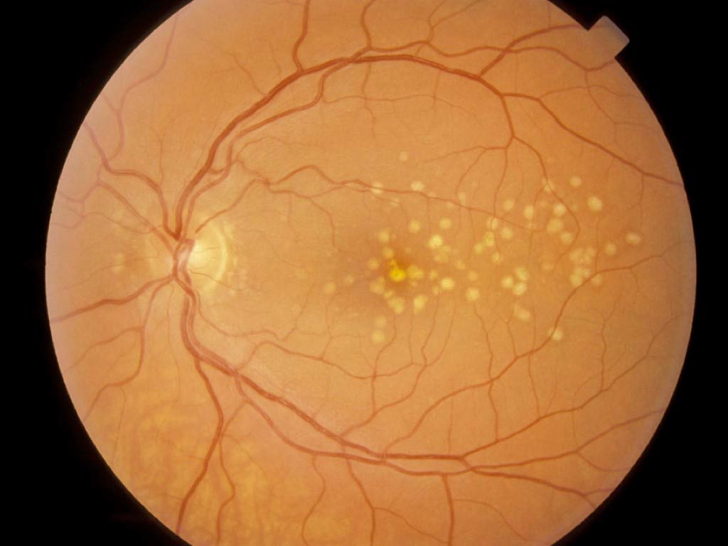
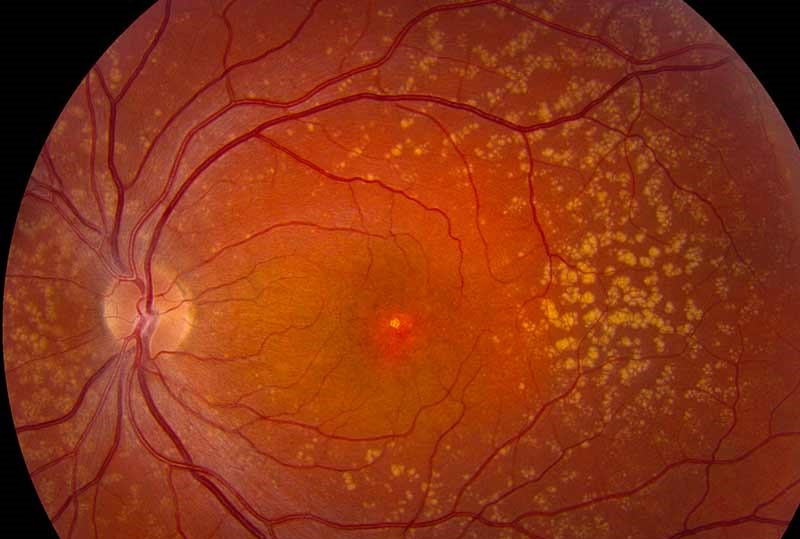
Hard drusen appear as small, round, yellow-white dots with crisp borders, and most adults over 50 have a few, which count as normal aging changes. On OCT, they produce small, smooth RPE elevations with homogeneous content, although very numerous small drusen deserve closer review, because some of these eyes progress.
Soft Drusen: Large, Blurred Borders
Soft drusen are larger and pale yellow with indistinct margins, and they cluster in the central macula and may coalesce into confluent sheets. On OCT, they form dome-shaped RPE elevations with medium reflective content, and confluent soft drusen can evolve into a drusenoid pigment epithelial detachment.
Soft Drusen and AMD Risk
Large drusen define intermediate AMD when no late features exist, and in the AREDS simplified scale, each eye with large drusen adds one risk point. Pigment abnormalities add another point per eye, and the total estimates five-year risk of late AMD, while soft drusen versus hard drusen offers a side-by-side comparison.
Cuticular Drusen Among Drusen Types
Cuticular drusen, also called basal laminar drusen, appear as numerous small, uniform yellow dots, and patients often present younger than in typical AMD. Studies link this phenotype to complement factor H gene variants, and our basal laminar drusen report covers the genetics in more detail.
Cuticular Drusen on Angiography and OCT
Fluorescein angiography shows a “stars in the sky” pattern of early punctate hyperfluorescence, while on OCT the RPE takes a saw-tooth profile of small triangular elevations. However, these eyes can develop vitelliform lesions, geographic atrophy or macular neovascularization, so plan long-term monitoring despite the small lesion size.
Calcified Drusen Types and Atrophy Risk
Calcified drusen look glistening or refractile on fundus examination, and they often mark regressing drusen. On OCT, they show heterogeneous internal reflectivity, with hyporeflective cores and hyperreflective caps, and research links calcified nodules within drusen to progression toward geographic atrophy.
Pseudodrusen and Other Drusen Types
Reticular pseudodrusen, also called subretinal drusenoid deposits, form an interlacing network of pale dots, and they favor the superior macula and often extend toward the arcades. Because they sit above the RPE, they behave differently from classic drusen, and their presence raises the risk of geographic atrophy and type 3 macular neovascularization.
Imaging Reticular Pseudodrusen
Color photos often miss them, whereas infrared reflectance and blue light autofluorescence show the network more clearly. On OCT, they appear as hyperreflective mounds or cones above the RPE, sometimes breaching the ellipsoid zone, while reticular pseudodrusen updates covers current concepts.
Multimodal Imaging for Drusen Types
First, use the fundus exam and color photo to judge size and borders. Next, use OCT to locate the deposit relative to the RPE. Finally, add infrared, autofluorescence or angiography when cuticular drusen or pseudodrusen seem likely.
Yellow Spots That Mimic Drusen
- Hard exudates, which sit in the outer plexiform layer and often ring a leak
- Optic disc drusen, which lie in the nerve head, away from the macula
- Pattern dystrophy and adult vitelliform lesions
- Stargardt flecks, especially in children and young adults
Counseling Patients About Drusen Types
Explain which type the patient has and what it means for that eye. For example, a few hard drusen usually need routine checks only, while large soft drusen justify AREDS2 supplements and home Amsler grid monitoring. Advise smokers to stop as well, because smoking raises the risk of progression.
Follow-Up for Drusen Types
Repeat OCT at intervals that match each patient’s risk profile, and watch for new hyperreflective foci, drusen collapse, hyporeflective cores and outer retinal thinning. Ask about new distortion at every visit, and then image within days when sudden symptoms suggest neovascularization.
Pitfalls to Avoid
- Do not label every yellow dot as drusen without OCT confirmation
- Never ignore pseudodrusen because color photos look nearly normal
- Do not dismiss cuticular drusen in a young patient, since they can progress
- Remember that collapsing drusen may herald atrophy


Document what you see
Two smartphone imaging tools built for everyday clinic use — one for the slit lamp, one for the fundus.
From Choroida — the team behind this siteReferences
- Ferris FL 3rd, Wilkinson CP, Bird A, et al. Clinical classification of age-related macular degeneration. Ophthalmology. 2013;120:844-851.
- Ferris FL, Davis MD, Clemons TE, et al. A simplified severity scale for age-related macular degeneration: AREDS report no. 18. Arch Ophthalmol. 2005;123:1570-1574.
- Spaide RF, Curcio CA. Drusen characterization with multimodal imaging. Retina. 2010;30:1441-1454.
- Balaratnasingam C, Cherepanoff S, Dolz-Marco R, et al. Cuticular drusen: clinical phenotypes and natural history defined using multimodal imaging. Ophthalmology. 2018;125:100-118.
- Tan ACS, Pilgrim MG, Fearn S, et al. Calcified nodules in retinal drusen are associated with disease progression in age-related macular degeneration. Sci Transl Med. 2018;10:eaat4544.

Writes and reviews the clinical articles on Educate. Every post is checked against peer-reviewed sources before it goes live.