AV ratio describes the width of a retinal arteriole relative to its paired venule.
Clinicians estimate it near the optic disc during routine fundus examination, and in healthy eyes, arterioles look about two thirds as wide as venules. However, bedside estimates vary widely between observers.
What the AV Ratio Measures
The ratio compares the visible blood columns of an arteriole and a neighboring venule, and a healthy vessel wall is transparent, so the examiner sees only the red cell column.
Therefore, wall thickening can change the reading before the lumen truly narrows. The ratio also reflects both vessels at once, which complicates interpretation.
Normal AV Ratio Values
Most clinical texts quote a normal ratio of about 2:3, which is roughly 0.67 in decimal terms. Small differences between quadrants and between eyes are common. As a result, a single borderline reading rarely means disease.
Fundus Explorer Pro
Photograph the retinal findings described here with the phone already in your pocket — 22 D optics and built-in illumination in one handheld unit.
From Choroida — the team behind this siteTelling Arterioles From Venules
Arterioles appear lighter red, narrower and straighter, and they also show a brighter central light reflex. Venules, by contrast, look darker and wider, with a duller reflex. In addition, arterioles usually cross venules, while two vessels of the same type seldom cross.
Bedside Estimation of the AV Ratio
- Dilate the pupil and focus carefully on the vessel edges
- Choose an arteriole and venule running together in the same quadrant
- Measure about half to one disc diameter from the disc margin
- Compare blood column widths, ignoring the light reflex
- Repeat in the other quadrants, then record the typical value
Choosing the Instrument
The direct ophthalmoscope gives high magnification and suits caliber comparison. However, its small field limits quadrant-by-quadrant review, while a 78 or 90 diopter lens at the slit lamp adds stereopsis and a wider view.
Generalized Arteriolar Narrowing
Chronic hypertension narrows arterioles diffusely through vasoconstriction and later sclerosis. As a result, the ratio drops toward 1:2, 1:3 or lower.
Keith, Wagener and Barker treated narrowing as an early grade in their classic scheme. Wong and Mitchell later grouped generalized narrowing with mild hypertensive retinopathy.
AV Ratio in Hypertensive Retinopathy
A low ratio supports chronic hypertension, but it does not prove it. Therefore, look for companion signs such as arteriovenous nicking, copper wiring and flame hemorrhages.
Cotton wool spots point to moderate retinopathy. Disc edema with these signs suggests malignant hypertension and needs same-day blood pressure assessment.
AV Ratio in Retinal Vein Occlusion
Vein occlusion lowers the ratio from the venous side. In central retinal vein occlusion, every quadrant shows dilated, tortuous veins. Branch occlusion, by contrast, changes only the affected sector, and in that case, the unaffected quadrant serves as an internal control.
Changes After Artery Occlusion
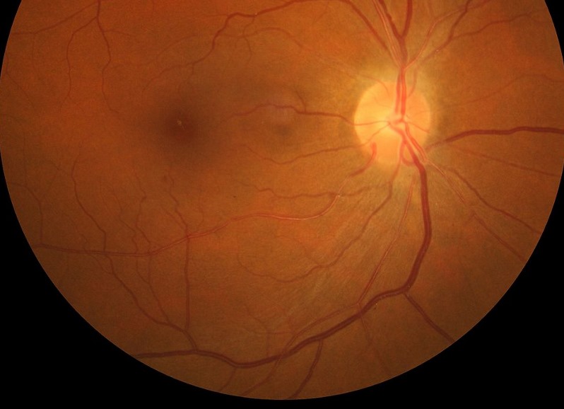
Artery occlusion lowers the ratio from the arteriolar side. Arterioles look attenuated, and the blood column may segment into boxcars.
In branch retinal artery occlusion, the change follows the occluded territory. Consequently, two eyes with similar ratios may reflect opposite mechanisms.
Venular Widening
Population studies link wider venules with smoking, inflammation, obesity and diabetes. Because the AV ratio merges both calibers, venular widening can mimic arteriolar narrowing. For this reason, research studies now report arteriolar and venular calibers separately.
Quantitative AV Ratio Measurement
The ARIC investigators measured vessels on digitized photographs in a defined zone around the disc and then summarized the data as central retinal arteriolar and venular equivalents. Knudtson and colleagues later revised the formulas using the six largest vessels of each type.
Cardiovascular Meaning
ARIC linked retinal microvascular abnormalities with incident stroke, independent of traditional risk factors. Similarly, the Rotterdam Study tied arteriolar narrowing to blood pressure and venular widening to inflammatory markers. However, these associations describe populations and do not predict an individual patient’s risk (see retinal vessel caliber and cardiovascular risk).
Factors That Distort the Reading
Several conditions shift the apparent caliber, and refractive error changes image magnification, although the ratio partly cancels this effect. Arteriolosclerosis also broadens the light reflex and makes the column look narrower. In addition, poor focus, media opacity and venous pulsation all reduce precision.
Counseling the Patient
Explain first that narrowed arterioles reflect long-standing blood pressure effects on small vessels. The finding rarely threatens vision by itself.
However, it justifies referral for blood pressure assessment and cardiovascular risk review. Better control may slow further change, although established sclerosis may persist.
Pitfalls to Avoid
- Do not measure the light reflex instead of the blood column
- Avoid reading the ratio on the disc, because vessels branch there
- Never call a low ratio hypertensive before checking the veins
- Do not compare an arteriole with a venule from another quadrant
- Avoid relying on one segment, because focal narrowing varies along the vessel


Document what you see
Two smartphone imaging tools built for everyday clinic use — one for the slit lamp, one for the fundus.
From Choroida — the team behind this siteReferences
- Wong TY, Mitchell P. Hypertensive retinopathy. N Engl J Med. 2004;351:2310-2317.
- Hubbard LD, Brothers RJ, King WN, et al. Methods for evaluation of retinal microvascular abnormalities associated with hypertension/sclerosis in the Atherosclerosis Risk in Communities Study. Ophthalmology. 1999;106:2269-2280.
- Knudtson MD, Lee KE, Hubbard LD, et al. Revised formulas for summarizing retinal vessel diameters. Curr Eye Res. 2003;27:143-149.
- Wong TY, Klein R, Couper DJ, et al. Retinal microvascular abnormalities and incident stroke: the Atherosclerosis Risk in Communities Study. Lancet. 2001;358:1134-1140.
- Ikram MK, de Jong FJ, Vingerling JR, et al. Are retinal arteriolar or venular diameters associated with markers for cardiovascular disorders? The Rotterdam Study. Invest Ophthalmol Vis Sci. 2004;45:2129-2134.
Test yourself
A few questions straight from this article.
-
What normal retinal arteriole-to-venule (AV) ratio do most clinical texts quote?
Most texts quote a normal AV ratio of about 2:3, roughly 0.67, meaning arterioles look about two thirds as wide as venules. -
When estimating the retinal AV ratio, what does the examiner actually see in a healthy vessel?
A healthy vessel wall is transparent, so only the blood column is visible; wall thickening can therefore change the reading before the lumen narrows. -
Which fundus feature identifies a retinal arteriole rather than a venule?
Arterioles look lighter red, narrower and straighter with a brighter central reflex, while venules are darker and wider with a duller reflex. -
Where should a bedside retinal AV ratio be estimated?
Measure about half to one disc diameter from the disc margin; reading on the disc is avoided because vessels branch there. -
In chronic hypertension with generalized arteriolar narrowing, the retinal AV ratio falls toward which values?
Chronic hypertension narrows arterioles diffusely through vasoconstriction and later sclerosis, so the ratio drops toward 1:2, 1:3 or lower. -
In a patient with a low AV ratio and hypertensive retinal signs, which added finding suggests malignant hypertension?
Disc edema alongside hypertensive signs suggests malignant hypertension and needs same-day blood pressure assessment. -
In branch retinal vein occlusion, what can serve as an internal control when judging the AV ratio?
Branch occlusion changes only the affected sector, unlike central occlusion where every quadrant shows dilated veins, so an unaffected quadrant acts as a control. -
Which arteriolar finding accompanies a reduced AV ratio after retinal artery occlusion?
Artery occlusion lowers the ratio from the arteriolar side: arterioles look attenuated and the blood column may segment into boxcars. -
Why do research studies now report retinal arteriolar and venular calibres separately rather than as an AV ratio?
Wider venules are linked with smoking, inflammation, obesity and diabetes, and because the ratio merges both calibres they can mimic arteriolar narrowing. -
How does arteriolosclerosis distort the apparent retinal arteriolar calibre?
Arteriolosclerosis broadens the central light reflex, which makes the blood column look narrower than it is.

Writes and reviews the clinical articles on Educate. Every post is checked against peer-reviewed sources before it goes live.