RNFL defect is a localized or diffuse loss of the retinal nerve fiber layer seen on fundus examination.
In glaucoma, it often appears before reproducible visual field loss, so the sign rewards careful observation at the slit lamp and on photographs.
Normal Nerve Fiber Pattern
Healthy axons create fine silver striations that radiate from the disc, and they look brightest along the superior and inferior arcades, where the layer is thickest. In addition, small retinal vessels appear partly buried in this reflective layer, while the nasal and papillomacular sectors look duller because the layer there is thinner.
What an RNFL Defect Looks Like
A localized defect is a dark band that interrupts the normal striations, and its edges are usually sharp, while the band widens as it leaves the disc. Inside it, the retinal surface looks matte and slightly darker, so vessels crossing the band look sharply outlined and exposed.
Fundus Explorer Pro
Photograph the retinal findings described here with the phone already in your pocket — 22 D optics and built-in illumination in one handheld unit.
From Choroida — the team behind this siteWedge-Shaped RNFL Defect
The classic wedge points its apex toward the disc margin and then fans out along the arcuate course of the fibers. Most wedges sit inferotemporally, followed by the superotemporal sector, and Jonas and Schiro found these defects commonly in glaucoma and uncommonly in normal eyes.
Diffuse RNFL Defect
Diffuse loss is harder to see because it has no sharp border. Instead, the whole arcade loses brightness and texture, and vessels then look bare across a wide area. Comparing both eyes, and the superior with the inferior arcade, helps because asymmetry draws attention.
Seeing the RNFL Defect Clinically
Use a dilated pupil, a 78 D or 90 D lens, and the green filter, and first scan along each arcade, then return to the disc margin. Young, pigmented fundi show fibers well, while older and lightly pigmented eyes show them poorly.
Red-Free Photography for RNFL Defect
Green light raises contrast between reflective fibers and the darker defect, so red-free photos are the classic way to document the sign (see red-free photography). Wide images of both arcades allow side-to-side comparison, and serial photos with identical settings reveal widening over time.
Why the Sign Appears
Glaucomatous injury damages ganglion cell axons at the lamina cribrosa. Because axons travel in arcuate bundles, focal damage at the rim removes a whole bundle, and the corresponding wedge then becomes visible downstream. Quigley and Addicks correlated photographic defects with histologic axon loss.
RNFL Defect and Disc Hemorrhage
A splinter hemorrhage at the rim often marks the site of a developing wedge, and Airaksinen and colleagues reported that hemorrhages preceded nerve fiber defects in ocular hypertension. Therefore, inspect the arcade next to any hemorrhage (see optic disc hemorrhage).
RNFL Defect and Field Loss
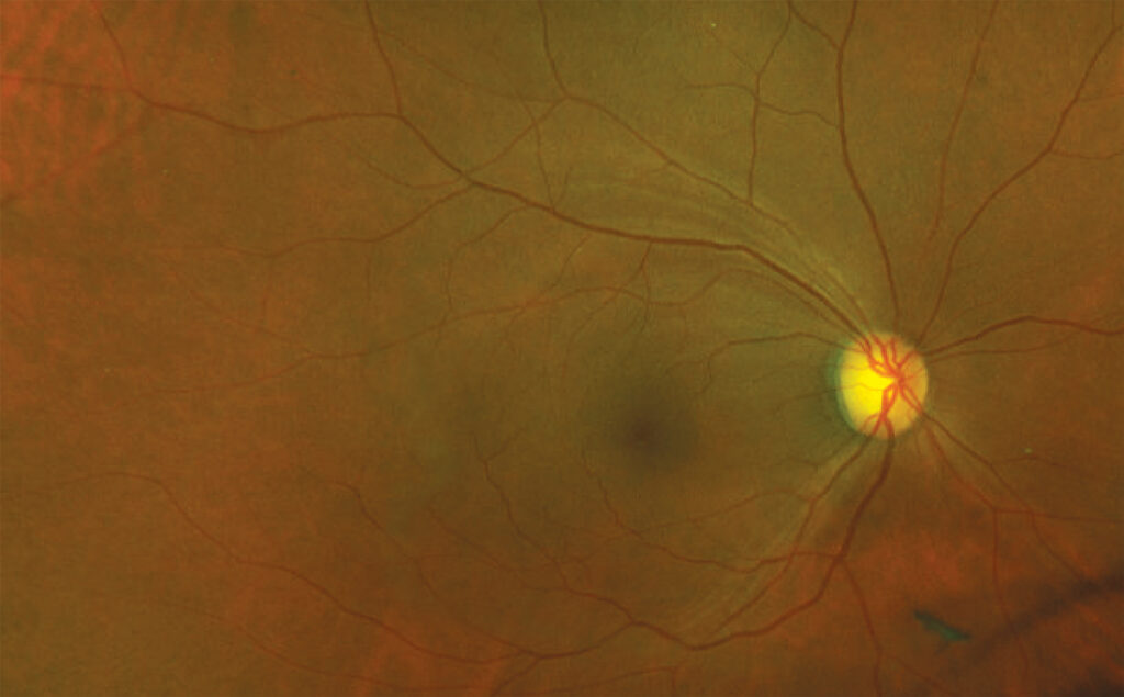
An inferior wedge predicts a superior arcuate defect or nasal step, and similarly, a superior wedge predicts an inferior field defect. Sommer and colleagues showed that nerve fiber atrophy often precedes detectable field loss by years, so a clear wedge with a normal field may represent preperimetric glaucoma.
Mimics of RNFL Defect
- Slit-like pseudodefects: narrow, thinner than a vessel, and stop short of the disc
- Old cotton-wool spots: leave focal defects in diabetes or hypertension
- Branch retinal artery occlusion: sectoral loss matching the occluded territory
- Myelinated fibers: bright patches make adjacent normal retina look dark (see myelinated nerve fibers)
- Optic disc drusen, prior optic neuritis, and ischemic optic neuropathy
- High myopia: tessellation and peripapillary atrophy hide fiber texture
OCT Correlation
OCT measures peripapillary thickness and flags thin sectors. However, a narrow wedge can fall between sectors and still look normal on the circle scan. Deviation maps from wider scans display wedges more clearly, so photos and OCT complement each other.
Management When You Find an RNFL Defect
A defect alone does not warrant treatment, so first check pressure, gonioscopy, corneal thickness, and fields. Then decide on the full risk profile and any documented progression, and when the pattern does not fit glaucoma, look for a nonglaucomatous cause.
Follow-Up
Repeat photography and OCT at intervals set by risk, where widening, deepening, or a new wedge signals progression, and record any new hemorrhage at the same location.
Counseling the Patient
Explain that the photo shows a small area of lost nerve fibers, and because many patients fear sudden blindness, describe their actual field status. Lost fibers do not regrow, while treatment aims to protect the remaining ones.
Pitfalls to Avoid
- Do not call a narrow slit a true defect
- Avoid judging fibers through a small pupil or dense cataract
- Do not dismiss a wedge because the field is normal
- Consider nonglaucomatous causes when the rim looks healthy


Document what you see
Two smartphone imaging tools built for everyday clinic use — one for the slit lamp, one for the fundus.
From Choroida — the team behind this siteReferences
- Hoyt WF, Frisén L, Newman NM. Fundoscopy of nerve fiber layer defects in glaucoma. Invest Ophthalmol. 1973;12:814-829.
- Sommer A, Katz J, Quigley HA, et al. Clinically detectable nerve fiber atrophy precedes the onset of glaucomatous field loss. Arch Ophthalmol. 1991;109:77-83.
- Jonas JB, Schiro D. Localised wedge shaped defects of the retinal nerve fibre layer in glaucoma. Br J Ophthalmol. 1994;78:285-290.
- Airaksinen PJ, Mustonen E, Alanko HI. Optic disc hemorrhages precede retinal nerve fiber layer defects in ocular hypertension. Acta Ophthalmol (Copenh). 1981;59:627-641.
- Quigley HA, Addicks EM. Quantitative studies of retinal nerve fiber layer defects. Arch Ophthalmol. 1982;100:807-814.

Writes and reviews the clinical articles on Educate. Every post is checked against peer-reviewed sources before it goes live.